 Movie
Movie Controller


+ Open data

- Basic information
| Entry | Database: PDB / ID: 5h72 | ||||||
|---|---|---|---|---|---|---|---|
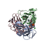
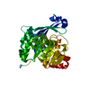
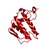
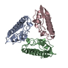
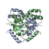
| Title | Structure of the periplasmic domain of FliP | ||||||
 Components Components | Flagellar biosynthetic protein FliP | ||||||
 Keywords Keywords |  BIOSYNTHETIC PROTEIN / Flagellar protein export BIOSYNTHETIC PROTEIN / Flagellar protein export | ||||||
| Function / homology |  Function and homology information Function and homology informationbacterial-type flagellum organization / bacterial-type flagellum basal body / protein secretion / membrane => GO:0016020 / plasma membraneSimilarity search - Function | ||||||
| Biological species |   Thermotoga maritima MSB8 (bacteria) Thermotoga maritima MSB8 (bacteria) | ||||||
| Method | X-RAY DIFFRACTION / SYNCHROTRON / SAD / Resolution: 2.4 Å | ||||||
 Authors Authors | Fukumura, T. / Kawaguchi, T. / Saijo-Hamano, Y. / Namba, K. / Minamino, T. / Imada, K. | ||||||
 Citation Citation | Journal: PLoS Biol. / Year: 2017 Title: Assembly and stoichiometry of the core structure of the bacterial flagellar type III export gate complex Authors: Fukumura, T. / Makino, F. / Dietsche, T. / Kinoshita, M. / Kato, T. / Wagner, S. / Namba, K. / Imada, K. / Minamino, T. #1: Journal: Acta Crystallogr.,Sect.F / Year: 2014 Title: Crystallization and preliminary X-ray analysis of the periplasmic domain of FliP, an integral membrane component of the bacterial flagellar type III protein-export apparatus Authors: Fukumura, T. / Furukawa, Y. / Kawaguchi, T. / Saijo-Hamano, Y. / Namba, K. / Imada, K. / Minamino, T. | ||||||
| History |
|
- Structure visualization
| Structure viewer | Molecule: MolmilJmol/JSmol |
|---|
- Downloads & links
-Download
| PDBx/mmCIF format | 5h72.cif.gz | 125.5 KB | Display | PDBx/mmCIF format |
|---|---|---|---|---|
| PDB format | pdb5h72.ent.gz | 100.6 KB | Display | PDB format |
| PDBx/mmJSON format | 5h72.json.gz | Tree view | PDBx/mmJSON format | |
| Others |  Other downloads Other downloads |
-Validation report
| Arichive directory | https://data.pdbj.org/pub/pdb/validation_reports/h7/5h72ftp://data.pdbj.org/pub/pdb/validation_reports/h7/5h72 | HTTPS FTP |
|---|
-Related structure data
| Similar structure data |
|---|


-Links
 PDBj
PDBj
- Assembly
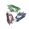
| Deposited unit | 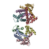
| ||||||||
|---|---|---|---|---|---|---|---|---|---|
| 1 | 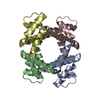
| ||||||||
| 2 | 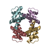
| ||||||||
| Unit cell |
| ||||||||
| Components on special symmetry positions |
|
-Components
| #1: Protein | Mass: 9433.692 Da / Num. of mol.: 8 / Fragment: periplasmic fragment, UNP residues 110-188 Source method: isolated from a genetically manipulated source Source: (gene. exp.) Thermotoga maritima MSB8 (bacteria) / Strain: MSB8 / Gene: fliP, TM_0698, Tmari_0698 / Production host: Escherichia coli (E. coli) / References: UniProt: Q9WZG2#2: Water | ChemComp-HOH / | Water Mass: 18.015 Da / Num. of mol.: 393 / Source method: isolated from a natural source / Formula: H2O Mass: 18.015 Da / Num. of mol.: 393 / Source method: isolated from a natural source / Formula: H2O |
|---|
-Experimental details
-Experiment
| Experiment | Method: X-RAY DIFFRACTION / Number of used crystals: 1 |
|---|
- Sample preparation
| Crystal | Density Matthews: 2.45 Å3/Da / Density % sol: 49.7 % |
|---|---|
| Crystal grow | Temperature: 277 K / Method: vapor diffusion, hanging drop / pH: 4.4 / Details: 0.1M phosphate-citrate pH 4.4, 36% MPD |
-Data collection
| Diffraction | Mean temperature: 100 K |
|---|---|
| Diffraction source | Source: SYNCHROTRON / Site: SPring-8  / Beamline: BL41XU / Wavelength: 1 Å / Beamline: BL41XU / Wavelength: 1 Å |
| Detector | Type: RAYONIX MX225HE / Detector: CCD / Date: Dec 2, 2012 |
| Radiation | Monochromator: Double-crystal monochromator / Protocol: SINGLE WAVELENGTH / Monochromatic (M) / Laue (L): M / Scattering type: x-ray |
| Radiation wavelength | Wavelength: 1 Å / Relative weight: 1 |
| Reflection | Resolution: 2.4→43.6 Å / Num. obs: 30262 / % possible obs: 99.9 % / Observed criterion σ(F): 0 / Observed criterion σ(I): 0 / Redundancy: 7.1 % / Biso Wilson estimate: 41 Å2 / Rmerge(I) obs: 0.088 / Net I/σ(I): 13.5 |
| Reflection shell | Resolution: 2.4→2.53 Å / Redundancy: 7.2 % / Rmerge(I) obs: 0.357 / Mean I/σ(I) obs: 5.5 / % possible all: 100 |
- Processing
| Software |
| ||||||||||||||||||||||||||||||||||||||||||||||||||||||||||||||||||||||||||||||||||||
|---|---|---|---|---|---|---|---|---|---|---|---|---|---|---|---|---|---|---|---|---|---|---|---|---|---|---|---|---|---|---|---|---|---|---|---|---|---|---|---|---|---|---|---|---|---|---|---|---|---|---|---|---|---|---|---|---|---|---|---|---|---|---|---|---|---|---|---|---|---|---|---|---|---|---|---|---|---|---|---|---|---|---|---|---|---|
| Refinement | Method to determine structure: SAD / Resolution: 2.4→42.923 Å / SU ML: 0.23 / Cross valid method: FREE R-VALUE / σ(F): 1.35 / Phase error: 31.63 / Stereochemistry target values: ML
| ||||||||||||||||||||||||||||||||||||||||||||||||||||||||||||||||||||||||||||||||||||
| Solvent computation | Shrinkage radii: 0.9 Å / VDW probe radii: 1.11 Å / Solvent model: FLAT BULK SOLVENT MODEL | ||||||||||||||||||||||||||||||||||||||||||||||||||||||||||||||||||||||||||||||||||||
| Refinement step | Cycle: LAST / Resolution: 2.4→42.923 Å
| ||||||||||||||||||||||||||||||||||||||||||||||||||||||||||||||||||||||||||||||||||||
| Refine LS restraints |
| ||||||||||||||||||||||||||||||||||||||||||||||||||||||||||||||||||||||||||||||||||||
| LS refinement shell |
|
