 Movie
Movie Controller

[English] 日本語
 Yorodumi
- PDB-5h4h: Structure of PIN-domain protein (VapC4 toxin) from Pyrococcus hor... -
+ Open data

- Basic information
| Entry | Database: PDB / ID: 5h4h | ||||||
|---|---|---|---|---|---|---|---|
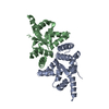
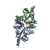
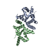
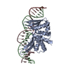
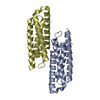
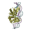
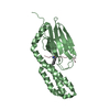
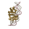
| Title | Structure of PIN-domain protein (VapC4 toxin) from Pyrococcus horikoshii determined at 2.2 A resolution | ||||||
 Components Components | Ribonuclease VapC4 | ||||||
 Keywords Keywords |  HYDROLASE / Pyrococcus horikoshii / hypothetical protein / PIN-domain HYDROLASE / Pyrococcus horikoshii / hypothetical protein / PIN-domain | ||||||
| Function / homology |  Function and homology information Function and homology informationRNA nuclease activity / Hydrolases; Acting on ester bonds / magnesium ion bindingSimilarity search - Function | ||||||
| Biological species |   Pyrococcus horikoshii OT3 (archaea) Pyrococcus horikoshii OT3 (archaea) | ||||||
| Method | X-RAY DIFFRACTION / MOLECULAR REPLACEMENT / Resolution: 2.23 Å | ||||||
 Authors Authors | Biswas, A. / Hatti, K. / Srinivasan, N. / Murthy, M.R.N. / Sekar, K. | ||||||
 Citation Citation | Journal: J. Struct. Biol. / Year: 2017 Title: Structure determination of contaminant proteins using the MarathonMR procedure Authors: Hatti, K. / Biswas, A. / Chaudhary, S. / Dadireddy, V. / Sekar, K. / Srinivasan, N. / Murthy, M.R.N. | ||||||
| History |
|
- Structure visualization
| Structure viewer | Molecule: MolmilJmol/JSmol |
|---|
- Downloads & links
-Download
| PDBx/mmCIF format | 5h4h.cif.gz | 69.1 KB | Display | PDBx/mmCIF format |
|---|---|---|---|---|
| PDB format | pdb5h4h.ent.gz | 50.6 KB | Display | PDB format |
| PDBx/mmJSON format | 5h4h.json.gz | Tree view | PDBx/mmJSON format | |
| Others |  Other downloads Other downloads |
-Validation report
| Arichive directory | https://data.pdbj.org/pub/pdb/validation_reports/h4/5h4hftp://data.pdbj.org/pub/pdb/validation_reports/h4/5h4h | HTTPS FTP |
|---|
-Related structure data
| Related structure data |  5h3lC  5h4fC  5h4gC  1v96S C: citing same article ( S: Starting model for refinement |
|---|---|
| Similar structure data |


-Links
 PDBj
PDBj

- Assembly
| Deposited unit | 
| ||||||||
|---|---|---|---|---|---|---|---|---|---|
| 1 |
| ||||||||
| Unit cell |
|
-Components
| #1: Protein | Mass: 17210.270 Da / Num. of mol.: 2 Source method: isolated from a genetically manipulated source Source: (gene. exp.) Pyrococcus horikoshii OT3 (archaea) / Strain: OT3 / Gene: vapC4, PH0500 / Production host:  Escherichia coli (E. coli) Escherichia coli (E. coli)References: UniProt: O58236, Hydrolases; Acting on ester bonds#2: Chemical |   Mass: 112.411 Da / Num. of mol.: 2 / Source method: obtained synthetically / Formula: Cd Mass: 112.411 Da / Num. of mol.: 2 / Source method: obtained synthetically / Formula: Cd#3: Water | ChemComp-HOH / | Water Mass: 18.015 Da / Num. of mol.: 19 / Source method: isolated from a natural source / Formula: H2O Mass: 18.015 Da / Num. of mol.: 19 / Source method: isolated from a natural source / Formula: H2O |
|---|
-Experimental details
-Experiment
| Experiment | Method: X-RAY DIFFRACTION / Number of used crystals: 1 |
|---|
- Sample preparation
| Crystal | Density Matthews: 1.77 Å3/Da / Density % sol: 30.58 % |
|---|---|
| Crystal grow | Temperature: 295 K / Method: vapor diffusion, hanging drop / pH: 8.5 Details: 0.2M Sodium acetate trihydrate, 0.1M Tris-HCl, 30%(w/v) PEG 4000 |
-Data collection
| Diffraction | Mean temperature: 300 K |
|---|---|
| Diffraction source | Source: ROTATING ANODE / Type: RIGAKU ULTRAX 18 / Wavelength: 1.54179 Å |
| Detector | Type: MARMOSAIC 225 mm CCD / Detector: CCD / Date: Jan 10, 2011 |
| Radiation | Protocol: SINGLE WAVELENGTH / Monochromatic (M) / Laue (L): M / Scattering type: x-ray |
| Radiation wavelength | Wavelength: 1.54179 Å / Relative weight: 1 |
| Reflection | Resolution: 2.21→51.85 Å / Num. obs: 11886 / % possible obs: 97.2 % / Redundancy: 3.3 % / Net I/σ(I): 8.5 |
| Reflection shell | Resolution: 2.21→2.29 Å / Redundancy: 3 % / Rmerge(I) obs: 0.42 / Mean I/σ(I) obs: 2.4 / CC1/2: 0.54 / % possible all: 71.2 |
- Processing
| Software |
| ||||||||||||||||||||||||||||||||||||||||||||||||||||||||||||||||||||||||||||||||||||||||||||||||||||||||||||||||||||||||||||||||||||||||||||||||||||||||||||||||||||||||||||||||||||||
|---|---|---|---|---|---|---|---|---|---|---|---|---|---|---|---|---|---|---|---|---|---|---|---|---|---|---|---|---|---|---|---|---|---|---|---|---|---|---|---|---|---|---|---|---|---|---|---|---|---|---|---|---|---|---|---|---|---|---|---|---|---|---|---|---|---|---|---|---|---|---|---|---|---|---|---|---|---|---|---|---|---|---|---|---|---|---|---|---|---|---|---|---|---|---|---|---|---|---|---|---|---|---|---|---|---|---|---|---|---|---|---|---|---|---|---|---|---|---|---|---|---|---|---|---|---|---|---|---|---|---|---|---|---|---|---|---|---|---|---|---|---|---|---|---|---|---|---|---|---|---|---|---|---|---|---|---|---|---|---|---|---|---|---|---|---|---|---|---|---|---|---|---|---|---|---|---|---|---|---|---|---|---|---|
| Refinement | Method to determine structure: MOLECULAR REPLACEMENT Starting model: 1V96 Resolution: 2.23→51.85 Å / Cor.coef. Fo:Fc: 0.935 / Cor.coef. Fo:Fc free: 0.9 / SU B: 9.757 / SU ML: 0.234 / Cross valid method: THROUGHOUT / ESU R: 0.514 / ESU R Free: 0.268 / Details: HYDROGENS HAVE BEEN ADDED IN THE RIDING POSITIONS
| ||||||||||||||||||||||||||||||||||||||||||||||||||||||||||||||||||||||||||||||||||||||||||||||||||||||||||||||||||||||||||||||||||||||||||||||||||||||||||||||||||||||||||||||||||||||
| Solvent computation | Ion probe radii: 0.8 Å / Shrinkage radii: 0.8 Å / VDW probe radii: 1.2 Å | ||||||||||||||||||||||||||||||||||||||||||||||||||||||||||||||||||||||||||||||||||||||||||||||||||||||||||||||||||||||||||||||||||||||||||||||||||||||||||||||||||||||||||||||||||||||
| Displacement parameters | Biso mean: 28.159 Å2
| ||||||||||||||||||||||||||||||||||||||||||||||||||||||||||||||||||||||||||||||||||||||||||||||||||||||||||||||||||||||||||||||||||||||||||||||||||||||||||||||||||||||||||||||||||||||
| Refinement step | Cycle: 1 / Resolution: 2.23→51.85 Å
| ||||||||||||||||||||||||||||||||||||||||||||||||||||||||||||||||||||||||||||||||||||||||||||||||||||||||||||||||||||||||||||||||||||||||||||||||||||||||||||||||||||||||||||||||||||||
| Refine LS restraints |
|