 Movie
Movie Controller


+ Open data

- Basic information
| Entry | Database: PDB / ID: 5gwo | ||||||
|---|---|---|---|---|---|---|---|
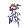
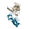
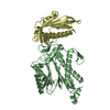
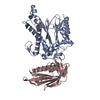
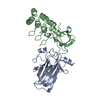
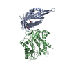
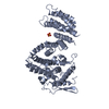
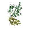
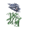
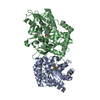
| Title | Crystal structure of RCAR3:PP2C S265F/I267M with (+)-ABA | ||||||
 Components Components |
| ||||||
 Keywords Keywords | HYDROLASE/RECEPTOR /  abscisic acid / ABA receptor / PP2C / HYDROLASE-RECEPTOR complex abscisic acid / ABA receptor / PP2C / HYDROLASE-RECEPTOR complex | ||||||
| Function / homology |  Function and homology information Function and homology informationnegative regulation of protein serine/threonine phosphatase activity / seed germination / response to water deprivation / abscisic acid binding / abscisic acid-activated signaling pathway / protein phosphatase inhibitor activity / myosin phosphatase activity / protein serine/threonine phosphatase activity / protein-serine/threonine phosphatase / response to cold ...negative regulation of protein serine/threonine phosphatase activity / seed germination / response to water deprivation / abscisic acid binding / abscisic acid-activated signaling pathway / protein phosphatase inhibitor activity / myosin phosphatase activity / protein serine/threonine phosphatase activity / protein-serine/threonine phosphatase / response to cold / signaling receptor activity / metal ion binding / nucleus / cytosol / cytoplasmSimilarity search - Function | ||||||
| Biological species |  Oryza sativa subsp. japonica (Japanese rice)Oryza sativa (Asian cultivated rice) Oryza sativa subsp. japonica (Japanese rice)Oryza sativa (Asian cultivated rice) | ||||||
| Method | X-RAY DIFFRACTION / SYNCHROTRON / MOLECULAR REPLACEMENT / Resolution: 2.816 Å | ||||||
 Authors Authors | Han, S. / Lee, S. | ||||||
 Citation Citation | Journal: Mol Plant / Year: 2017 Title: Modulation of ABA Signaling by Altering VxG Phi L Motif of PP2Cs in Oryza sativa. Authors: Han, S. / Min, M.K. / Lee, S.Y. / Lim, C.W. / Bhatnagar, N. / Lee, Y. / Shin, D. / Chung, K.Y. / Lee, S.C. / Kim, B.G. / Lee, S. | ||||||
| History |
|
- Structure visualization
| Structure viewer | Molecule: MolmilJmol/JSmol |
|---|
- Downloads & links
-Download
| PDBx/mmCIF format | 5gwo.cif.gz | 387.3 KB | Display | PDBx/mmCIF format |
|---|---|---|---|---|
| PDB format | pdb5gwo.ent.gz | 315.7 KB | Display | PDB format |
| PDBx/mmJSON format | 5gwo.json.gz | Tree view | PDBx/mmJSON format | |
| Others |  Other downloads Other downloads |
-Validation report
| Arichive directory | https://data.pdbj.org/pub/pdb/validation_reports/gw/5gwoftp://data.pdbj.org/pub/pdb/validation_reports/gw/5gwo | HTTPS FTP |
|---|
-Related structure data
| Related structure data | 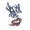 5gwpC 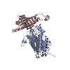 3rt0S C: citing same article ( S: Starting model for refinement |
|---|---|
| Similar structure data |
-Links
 PDBj
PDBj- Assembly
| Deposited unit | 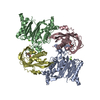
| ||||||||
|---|---|---|---|---|---|---|---|---|---|
| 1 | 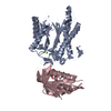
| ||||||||
| 2 | 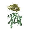
| ||||||||
| Unit cell |
|
-Components
| #1: Protein | Probability / OsPP2C50 Mass: 35585.645 Da / Num. of mol.: 2 / Fragment: UNP residues 59-385 / Mutation: E139A/E140A/K142A/S265F/I267M Source method: isolated from a genetically manipulated source Source: (gene. exp.) Oryza sativa subsp. japonica (Japanese rice)Gene: Os05g0537400, LOC_Os05g46040, OJ1741_B01.18, OSJNBa0052K01.2 Production host:  Escherichia coli (E. coli) Escherichia coli (E. coli)References: UniProt: Q6L5H6, protein-serine/threonine phosphatase #2: Protein | Mass: 20060.865 Da / Num. of mol.: 2 / Fragment: UNP residues 30-204 Source method: isolated from a genetically manipulated source Source: (gene. exp.) Oryza sativa (Asian cultivated rice) / Production host: Escherichia coli (E. coli) / References: UniProt: K4N2F7, UniProt: Q6EN42*PLUS#3: Chemical | ChemComp-MG /   Mass: 24.305 Da / Num. of mol.: 5 / Source method: obtained synthetically / Formula: Mg Mass: 24.305 Da / Num. of mol.: 5 / Source method: obtained synthetically / Formula: Mg#4: Chemical | Abscisic acid  Mass: 264.317 Da / Num. of mol.: 2 / Source method: obtained synthetically / Formula: C15H20O4 / Comment: hormone*YM Mass: 264.317 Da / Num. of mol.: 2 / Source method: obtained synthetically / Formula: C15H20O4 / Comment: hormone*YM#5: Water | ChemComp-HOH / | Water Mass: 18.015 Da / Num. of mol.: 37 / Source method: isolated from a natural source / Formula: H2O Mass: 18.015 Da / Num. of mol.: 37 / Source method: isolated from a natural source / Formula: H2O |
|---|
-Experimental details
-Experiment
| Experiment | Method: X-RAY DIFFRACTION / Number of used crystals: 1 |
|---|
- Sample preparation
| Crystal | Density Matthews: 2.53 Å3/Da / Density % sol: 51.31 % |
|---|---|
| Crystal grow | Temperature: 295 K / Method: vapor diffusion, hanging drop / Details: PEG 8000, MES pH 6.0, magnesium acetate, glycerol |
-Data collection
| Diffraction | Mean temperature: 100 K |
|---|---|
| Diffraction source | Source: SYNCHROTRON / Site: PAL/PLS  / Beamline: 5C (4A) / Wavelength: 0.9795 Å / Beamline: 5C (4A) / Wavelength: 0.9795 Å |
| Detector | Type: ADSC QUANTUM 315r / Detector: CCD / Date: Mar 22, 2016 |
| Radiation | Protocol: SINGLE WAVELENGTH / Monochromatic (M) / Laue (L): M / Scattering type: x-ray |
| Radiation wavelength | Wavelength: 0.9795 Å / Relative weight: 1 |
| Reflection | Resolution: 2.816→37.729 Å / Num. obs: 25506 / % possible obs: 97 % / Redundancy: 3.2 % / CC1/2: 0.993 / Rmerge(I) obs: 0.1066 / Rsym value: 0.1274 / Net I/σ(I): 13.54 |
| Reflection shell | Resolution: 2.816→2.917 Å |
- Processing
| Software |
| |||||||||||||||||||||||||||||||||||||||||||||||||||||||||||||||||||||||||||||||||||||||||||||||||||||||||
|---|---|---|---|---|---|---|---|---|---|---|---|---|---|---|---|---|---|---|---|---|---|---|---|---|---|---|---|---|---|---|---|---|---|---|---|---|---|---|---|---|---|---|---|---|---|---|---|---|---|---|---|---|---|---|---|---|---|---|---|---|---|---|---|---|---|---|---|---|---|---|---|---|---|---|---|---|---|---|---|---|---|---|---|---|---|---|---|---|---|---|---|---|---|---|---|---|---|---|---|---|---|---|---|---|---|---|
| Refinement | Method to determine structure: MOLECULAR REPLACEMENT Starting model: 3RT0 Resolution: 2.816→37.729 Å / SU ML: 0.4 / Cross valid method: NONE / σ(F): 1.98 / Phase error: 27.74
| |||||||||||||||||||||||||||||||||||||||||||||||||||||||||||||||||||||||||||||||||||||||||||||||||||||||||
| Solvent computation | Shrinkage radii: 0.9 Å / VDW probe radii: 1.11 Å | |||||||||||||||||||||||||||||||||||||||||||||||||||||||||||||||||||||||||||||||||||||||||||||||||||||||||
| Refinement step | Cycle: LAST / Resolution: 2.816→37.729 Å
| |||||||||||||||||||||||||||||||||||||||||||||||||||||||||||||||||||||||||||||||||||||||||||||||||||||||||
| Refine LS restraints |
| |||||||||||||||||||||||||||||||||||||||||||||||||||||||||||||||||||||||||||||||||||||||||||||||||||||||||
| LS refinement shell |
| |||||||||||||||||||||||||||||||||||||||||||||||||||||||||||||||||||||||||||||||||||||||||||||||||||||||||
| Refinement TLS params. | Method: refined / Origin x: -37.4819 Å / Origin y: 26.4132 Å / Origin z: -5.9916 Å
| |||||||||||||||||||||||||||||||||||||||||||||||||||||||||||||||||||||||||||||||||||||||||||||||||||||||||
| Refinement TLS group | Selection details: all |
