 Movie
Movie Controller


+ Open data

- Basic information
| Entry | Database: PDB / ID: 5gu7 | ||||||
|---|---|---|---|---|---|---|---|
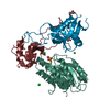
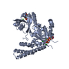
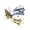
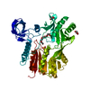
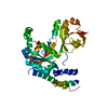
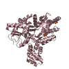
| Title | Crystal Structure of human ERp44 form II | ||||||
 Components Components | Endoplasmic reticulum resident protein 44 | ||||||
 Keywords Keywords |  CHAPERONE / quality control CHAPERONE / quality control | ||||||
| Function / homology |  Function and homology information Function and homology informationglycoprotein metabolic process / protein disulfide isomerase activity / response to unfolded protein / endoplasmic reticulum-Golgi intermediate compartment / response to endoplasmic reticulum stress / cell redox homeostasis / specific granule lumen / protein folding / endoplasmic reticulum lumen / Neutrophil degranulation ...glycoprotein metabolic process / protein disulfide isomerase activity / response to unfolded protein / endoplasmic reticulum-Golgi intermediate compartment / response to endoplasmic reticulum stress / cell redox homeostasis / specific granule lumen / protein folding / endoplasmic reticulum lumen / Neutrophil degranulation / endoplasmic reticulum membrane / cell surface / extracellular exosome / extracellular regionSimilarity search - Function | ||||||
| Biological species |  Homo sapiens (human) Homo sapiens (human) | ||||||
| Method | X-RAY DIFFRACTION / SYNCHROTRON / MOLECULAR REPLACEMENT / Resolution: 2.05 Å | ||||||
 Authors Authors | Watanabe, S. / Inaba, K. | ||||||
 Citation Citation | Journal: Proc. Natl. Acad. Sci. U.S.A. / Year: 2017 Title: Structural basis of pH-dependent client binding by ERp44, a key regulator of protein secretion at the ER-Golgi interface Authors: Watanabe, S. / Harayama, M. / Kanemura, S. / Sitia, R. / Inaba, K. | ||||||
| History |
|
- Structure visualization
| Structure viewer | Molecule: MolmilJmol/JSmol |
|---|
- Downloads & links
-Download
| PDBx/mmCIF format | 5gu7.cif.gz | 169.6 KB | Display | PDBx/mmCIF format |
|---|---|---|---|---|
| PDB format | pdb5gu7.ent.gz | 131.9 KB | Display | PDB format |
| PDBx/mmJSON format | 5gu7.json.gz | Tree view | PDBx/mmJSON format | |
| Others |  Other downloads Other downloads |
-Validation report
| Arichive directory | https://data.pdbj.org/pub/pdb/validation_reports/gu/5gu7ftp://data.pdbj.org/pub/pdb/validation_reports/gu/5gu7 | HTTPS FTP |
|---|
-Related structure data
| Related structure data |  5gu6C  2r2jS C: citing same article ( S: Starting model for refinement |
|---|---|
| Similar structure data |


-Links
 PDBj
PDBj


- Assembly
| Deposited unit | 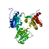
| ||||||||
|---|---|---|---|---|---|---|---|---|---|
| 1 |
| ||||||||
| 2 | 
| ||||||||
| Unit cell |
|
-Components
| #1: Protein | Mass: 45720.273 Da / Num. of mol.: 1 Source method: isolated from a genetically manipulated source Source: (gene. exp.) Homo sapiens (human) / Gene: ERP44, KIAA0573, TXNDC4, UNQ532/PRO1075 / Production host:  Escherichia coli (E. coli) / References: UniProt: Q9BS26 Escherichia coli (E. coli) / References: UniProt: Q9BS26 |
|---|---|
| #2: Water | ChemComp-HOH / Water Mass: 18.015 Da / Num. of mol.: 146 / Source method: isolated from a natural source / Formula: H2O Mass: 18.015 Da / Num. of mol.: 146 / Source method: isolated from a natural source / Formula: H2O |
-Experimental details
-Experiment
| Experiment | Method: X-RAY DIFFRACTION / Number of used crystals: 1 |
|---|
- Sample preparation
| Crystal | Density Matthews: 2.98 Å3/Da / Density % sol: 58.69 % |
|---|---|
| Crystal grow | Temperature: 277 K / Method: vapor diffusion, sitting drop / pH: 6.5 / Details: Na/K tartrate |
-Data collection
| Diffraction | Mean temperature: 100 K |
|---|---|
| Diffraction source | Source: SYNCHROTRON / Site: SPring-8  / Beamline: BL44XU / Wavelength: 0.9 Å / Beamline: BL44XU / Wavelength: 0.9 Å |
| Detector | Type: RAYONIX MX300HE / Detector: CCD / Date: Apr 18, 2015 |
| Radiation | Protocol: SINGLE WAVELENGTH / Monochromatic (M) / Laue (L): M / Scattering type: x-ray |
| Radiation wavelength | Wavelength: 0.9 Å / Relative weight: 1 |
| Reflection | Resolution: 2.05→37.913 Å / Num. obs: 34847 / % possible obs: 99.9 % / Redundancy: 17.1 % / CC1/2: 0.999 / Rmerge(I) obs: 0.063 / Net I/σ(I): 26 |
| Reflection shell | Resolution: 2.05→2.1 Å / Redundancy: 11.24 % / Rmerge(I) obs: 1.176 / Mean I/σ(I) obs: 2.3 / CC1/2: 0.764 / % possible all: 100 |
- Processing
| Software |
| ||||||||||||||||||||||||||||||||||||||||||||||||||||||||||||||||||||||||||||||||||||||||||||||||||||
|---|---|---|---|---|---|---|---|---|---|---|---|---|---|---|---|---|---|---|---|---|---|---|---|---|---|---|---|---|---|---|---|---|---|---|---|---|---|---|---|---|---|---|---|---|---|---|---|---|---|---|---|---|---|---|---|---|---|---|---|---|---|---|---|---|---|---|---|---|---|---|---|---|---|---|---|---|---|---|---|---|---|---|---|---|---|---|---|---|---|---|---|---|---|---|---|---|---|---|---|---|---|
| Refinement | Method to determine structure: MOLECULAR REPLACEMENT Starting model: 2R2J Resolution: 2.05→37.913 Å / SU ML: 0.25 / Cross valid method: FREE R-VALUE / σ(F): 1.34 / Phase error: 25.89
| ||||||||||||||||||||||||||||||||||||||||||||||||||||||||||||||||||||||||||||||||||||||||||||||||||||
| Solvent computation | Shrinkage radii: 0.9 Å / VDW probe radii: 1.11 Å | ||||||||||||||||||||||||||||||||||||||||||||||||||||||||||||||||||||||||||||||||||||||||||||||||||||
| Refinement step | Cycle: LAST / Resolution: 2.05→37.913 Å
| ||||||||||||||||||||||||||||||||||||||||||||||||||||||||||||||||||||||||||||||||||||||||||||||||||||
| Refine LS restraints |
| ||||||||||||||||||||||||||||||||||||||||||||||||||||||||||||||||||||||||||||||||||||||||||||||||||||
| LS refinement shell |
| ||||||||||||||||||||||||||||||||||||||||||||||||||||||||||||||||||||||||||||||||||||||||||||||||||||
| Refinement TLS params. | Method: refined / Refine-ID: X-RAY DIFFRACTION
| ||||||||||||||||||||||||||||||||||||||||||||||||||||||||||||||||||||||||||||||||||||||||||||||||||||
| Refinement TLS group |
|
