[English] 日本語
Yorodumi
- PDB-5gnb: Crystal Structure of the Receptor Binding Domain of the Spike Gly... -

+
Open data


ID or keywords:

Loading...

-
Basic information

Entry
Database: PDB / ID: 5gnb
Title
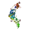
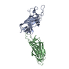
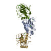
Crystal Structure of the Receptor Binding Domain of the Spike Glycoprotein of Human Betacoronavirus HKU1 (HKU1 1A-CTD, 2.3 angstrom, native-SAD phasing)
ComponentsSpike glycoproteinSpike protein
KeywordsVIRAL PROTEIN / Coronavirus spike protein / S1-CTD / receptor binding domain / receptor binding motif / virus entry / HKU1
Function / homology
Function and homology information


endocytosis involved in viral entry into host cell / host cell endoplasmic reticulum-Golgi intermediate compartment membrane / receptor-mediated virion attachment to host cell / fusion of virus membrane with host plasma membrane / fusion of virus membrane with host endosome membrane / viral envelope / host cell plasma membrane / virion membrane / membrane
Similarity search - Function
Spike (S) protein S1 subunit, N-terminal domain, murine hepatitis virus-like / Spike glycoprotein S2, coronavirus, C-terminal / Coronavirus spike glycoprotein S2, intravirion / Betacoronavirus spike (S) glycoprotein S1 subunit N-terminal (NTD) domain profile. / Spike glycoprotein, N-terminal domain superfamily / Betacoronavirus spike (S) glycoprotein S1 subunit C-terminal (CTD) domain profile. / Spike glycoprotein, betacoronavirus / Spike (S) protein S1 subunit, receptor-binding domain, betacoronavirus / Spike S1 subunit, receptor binding domain superfamily, betacoronavirus / Betacoronavirus spike glycoprotein S1, receptor binding ...Spike (S) protein S1 subunit, N-terminal domain, murine hepatitis virus-like / Spike glycoprotein S2, coronavirus, C-terminal / Coronavirus spike glycoprotein S2, intravirion / Betacoronavirus spike (S) glycoprotein S1 subunit N-terminal (NTD) domain profile. / Spike glycoprotein, N-terminal domain superfamily / Betacoronavirus spike (S) glycoprotein S1 subunit C-terminal (CTD) domain profile. / Spike glycoprotein, betacoronavirus / Spike (S) protein S1 subunit, receptor-binding domain, betacoronavirus / Spike S1 subunit, receptor binding domain superfamily, betacoronavirus / Betacoronavirus spike glycoprotein S1, receptor binding / Spike glycoprotein S1, N-terminal domain, betacoronavirus-like / Betacoronavirus-like spike glycoprotein S1, N-terminal / Spike glycoprotein S2, coronavirus, heptad repeat 1 / Spike glycoprotein S2, coronavirus, heptad repeat 2 / Coronavirus spike (S) glycoprotein S2 subunit heptad repeat 2 (HR2) region profile. / Coronavirus spike (S) glycoprotein S2 subunit heptad repeat 1 (HR1) region profile. / Spike glycoprotein S2 superfamily, coronavirus / Spike glycoprotein S2, coronavirus / Coronavirus spike glycoprotein S2 / Coronavirus spike glycoprotein S1, C-terminal / Coronavirus spike glycoprotein S1, C-terminal
Similarity search - Domain/homology
Biological speciesHuman coronavirus HKU1
MethodX-RAY DIFFRACTION / SYNCHROTRON / SAD / Resolution: 2.3 Å
AuthorsGuan, H. / Wojdyla, J.A. / Wang, M. / Cui, S.
CitationJournal: Nat Commun / Year: 2017
Title: Crystal structure of the receptor binding domain of the spike glycoprotein of human betacoronavirus HKU1
Authors: Ou, X. / Guan, H. / Qin, B. / Mu, Z. / Wojdyla, J.A. / Wang, M. / Dominguez, S.R. / Qian, Z. / Cui, S.
History
DepositionJul 20, 2016Deposition site: PDBJ / Processing site: PDBJ
Revision 1.0Jun 7, 2017Provider: repository / Type: Initial release
Revision 1.1Jun 14, 2017Group: Structure summary / Category: struct / struct_keywords / Item: _struct.title / _struct_keywords.text
Revision 1.2Sep 27, 2017Group: Data collection / Category: diffrn_detector / Item: _diffrn_detector.detector
Revision 1.3Jul 29, 2020Group: Data collection / Derived calculations / Structure summary
Category: chem_comp / entity ...chem_comp / entity / pdbx_chem_comp_identifier / pdbx_entity_nonpoly / struct_conn / struct_site / struct_site_gen
Item: _chem_comp.name / _chem_comp.type ..._chem_comp.name / _chem_comp.type / _entity.pdbx_description / _pdbx_entity_nonpoly.name / _struct_conn.pdbx_role
Description: Carbohydrate remediation / Provider: repository / Type: Remediation

-
Structure visualization

Structure viewerMolecule:
MolmilJmol/JSmol

Downloads & links

-
Assembly

Deposited unit
A: Spike glycoprotein
hetero molecules


Theoretical massNumber of molelcules
Total (without water)42,3105
Polymers41,4251
Non-polymers8854
Water4,216234
1


  • Idetical with deposited unit
  • defined by author
TypeNameSymmetry operationNumber
identity operation1_555x,y,z1
Buried area860 Å2
ΔGint12 kcal/mol
Surface area18850 Å2
Unit cell
Length a, b, c (Å)86.720, 183.990, 63.160
Angle α, β, γ (deg.)90.00, 90.00, 90.00
Int Tables number20
Space group name H-MC2221
Components on special symmetry positions
IDModelComponents
11A-980-

HOH

21A-1002-

HOH

-
Components

#1: Protein Spike glycoprotein / Spike protein / S glycoprotein / E2 / Peplomer protein


Mass: 41425.062 Da / Num. of mol.: 1 / Fragment: UNP residues 307-677
Source method: isolated from a genetically manipulated source
Source: (gene. exp.) Human coronavirus HKU1 (isolate N1) / Strain: isolate N1 / Gene: S, 3 / Production host: Trichoplusia ni (cabbage looper) / References: UniProt: Q5MQD0
#2: Sugar
ChemComp-NAG / 2-acetamido-2-deoxy-beta-D-glucopyranose / N-Acetylglucosamine


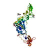
Type: D-saccharide, beta linking / Mass: 221.208 Da / Num. of mol.: 4
Source method: isolated from a genetically manipulated source
Formula: C8H15NO6
IdentifierTypeProgram
DGlcpNAcbCONDENSED IUPAC CARBOHYDRATE SYMBOLGMML 1.0
N-acetyl-b-D-glucopyranosamineCOMMON NAMEGMML 1.0
b-D-GlcpNAcIUPAC CARBOHYDRATE SYMBOLPDB-CARE 1.0
GlcNAcSNFG CARBOHYDRATE SYMBOLGMML 1.0
#3: Water ChemComp-HOH / water / Water


Mass: 18.015 Da / Num. of mol.: 234 / Source method: isolated from a natural source / Formula: H2O

-
Experimental details

-
Experiment

ExperimentMethod: X-RAY DIFFRACTION / Number of used crystals: 1

-
Sample preparation

CrystalDensity Matthews: 3.04 Å3/Da / Density % sol: 60 %
Description: THE ENTRY CONTAINS FRIEDEL PAIRS IN F_PLUS/MINUS COLUMNS.
Crystal growTemperature: 291 K / Method: vapor diffusion, hanging drop / pH: 8.5
Details: 0.1M Tris, 24%PEG4000, 0.2M MgCl2, 4% v/v Pentaerythritol ethoxylate (3/4 EO/OH)

-
Data collection

DiffractionMean temperature: 100 K
Diffraction sourceSource: SYNCHROTRON / Site: SLS / Beamline: X06DA / Wavelength: 2.08 Å
DetectorType: DECTRIS PILATUS 2M-F / Detector: PIXEL / Date: May 9, 2015
RadiationProtocol: SINGLE WAVELENGTH / Monochromatic (M) / Laue (L): M / Scattering type: x-ray
Radiation wavelengthWavelength: 2.08 Å / Relative weight: 1
ReflectionResolution: 2.3→49.2 Å / Num. obs: 43316 / % possible obs: 99.5 % / Observed criterion σ(I): -3 / Redundancy: 89.76 % / Biso Wilson estimate: 31.062 Å2 / CC1/2: 0.999 / Rmerge(I) obs: 0.23 / Net I/σ(I): 28.04
Reflection shellResolution: 2.3→2.36 Å / Redundancy: 30.92 % / Rmerge(I) obs: 1.28 / Mean I/σ(I) obs: 3.37 / CC1/2: 0.894 / % possible all: 93.9

-
Processing

Software
NameVersionClassification
PHENIX(1.10.1_2155: ???)refinement
XDSdata reduction
XDSdata scaling
SHELXCDphasing
RefinementMethod to determine structure: SAD / Resolution: 2.3→49.2 Å / SU ML: 0.26 / Cross valid method: THROUGHOUT / σ(F): 1.93 / Phase error: 23.97
Details: SF FILE CONTAINS FRIEDEL PAIRS UNDER I/F_MINUS AND I/F_PLUS COLUMNS.
RfactorNum. reflection% reflectionSelection details
Rfree0.2192 2155 4.98 %Random
Rwork0.1728 ---
obs0.1751 43282 99.43 %-
Solvent computationShrinkage radii: 0.9 Å / VDW probe radii: 1.11 Å
Displacement parametersBiso mean: 30.16 Å2
Refinement stepCycle: LAST / Resolution: 2.3→49.2 Å
ProteinNucleic acidLigandSolventTotal
Num. atoms2853 0 56 234 3143
Refine LS restraints
Refine-IDTypeDev idealNumber
X-RAY DIFFRACTIONf_bond_d0.0083070
X-RAY DIFFRACTIONf_angle_d0.8544213
X-RAY DIFFRACTIONf_dihedral_angle_d9.9681859
X-RAY DIFFRACTIONf_chiral_restr0.051468
X-RAY DIFFRACTIONf_plane_restr0.005546
LS refinement shell
Resolution (Å)Rfactor RfreeNum. reflection RfreeRfactor RworkNum. reflection RworkRefine-ID% reflection obs (%)
2.298-2.35150.30321370.23492595X-RAY DIFFRACTION93
2.3515-2.41030.25661400.21762720X-RAY DIFFRACTION99
2.4103-2.47540.23841450.20562765X-RAY DIFFRACTION100
2.4754-2.54830.28891410.18642762X-RAY DIFFRACTION100
2.5483-2.63050.29931400.18312734X-RAY DIFFRACTION100
2.6305-2.72450.21131480.17062760X-RAY DIFFRACTION100
2.7245-2.83360.22231470.17322745X-RAY DIFFRACTION100
2.8336-2.96250.23721440.18482731X-RAY DIFFRACTION100
2.9625-3.11870.23841440.19142764X-RAY DIFFRACTION100
3.1187-3.31410.23231430.17472762X-RAY DIFFRACTION100
3.3141-3.56990.18691470.16372755X-RAY DIFFRACTION100
3.5699-3.9290.20771480.15452774X-RAY DIFFRACTION100
3.929-4.49720.20861420.1342741X-RAY DIFFRACTION100
4.4972-5.66470.14631430.15082769X-RAY DIFFRACTION100
5.6647-49.20670.23021460.19472750X-RAY DIFFRACTION100

+
About Yorodumi

-
News

-
Feb 9, 2022. New format data for meta-information of EMDB entries

New format data for meta-information of EMDB entries

  • Version 3 of the EMDB header file is now the official format.
  • The previous official version 1.9 will be removed from the archive.

Related info.:EMDB header

External links:wwPDB to switch to version 3 of the EMDB data model

-
Aug 12, 2020. Covid-19 info

Covid-19 info

URL: https://pdbj.org/emnavi/covid19.php

New page: Covid-19 featured information page in EM Navigator.

Related info.:Covid-19 info / Mar 5, 2020. Novel coronavirus structure data

+
Mar 5, 2020. Novel coronavirus structure data

Novel coronavirus structure data

Related info.:Yorodumi Speices / Aug 12, 2020. Covid-19 info

External links:COVID-19 featured content - PDBj / Molecule of the Month (242):Coronavirus Proteases

+
Jan 31, 2019. EMDB accession codes are about to change! (news from PDBe EMDB page)

EMDB accession codes are about to change! (news from PDBe EMDB page)

  • The allocation of 4 digits for EMDB accession codes will soon come to an end. Whilst these codes will remain in use, new EMDB accession codes will include an additional digit and will expand incrementally as the available range of codes is exhausted. The current 4-digit format prefixed with “EMD-” (i.e. EMD-XXXX) will advance to a 5-digit format (i.e. EMD-XXXXX), and so on. It is currently estimated that the 4-digit codes will be depleted around Spring 2019, at which point the 5-digit format will come into force.
  • The EM Navigator/Yorodumi systems omit the EMD- prefix.

Related info.:Q: What is EMD? / ID/Accession-code notation in Yorodumi/EM Navigator

External links:EMDB Accession Codes are Changing Soon! / Contact to PDBj

+
Jul 12, 2017. Major update of PDB

Major update of PDB

  • wwPDB released updated PDB data conforming to the new PDBx/mmCIF dictionary.
  • This is a major update changing the version number from 4 to 5, and with Remediation, in which all the entries are updated.
  • In this update, many items about electron microscopy experimental information are reorganized (e.g. em_software).
  • Now, EM Navigator and Yorodumi are based on the updated data.

External links:wwPDB Remediation / Enriched Model Files Conforming to OneDep Data Standards Now Available in the PDB FTP Archive

-
Yorodumi

Thousand views of thousand structures

  • Yorodumi is a browser for structure data from EMDB, PDB, SASBDB, etc.
  • This page is also the successor to EM Navigator detail page, and also detail information page/front-end page for Omokage search.
  • The word "yorodu" (or yorozu) is an old Japanese word meaning "ten thousand". "mi" (miru) is to see.

Related info.:EMDB / PDB / SASBDB / Comparison of 3 databanks / Yorodumi Search / Aug 31, 2016. New EM Navigator & Yorodumi / Yorodumi Papers / Jmol/JSmol / Function and homology information / Changes in new EM Navigator and Yorodumi

Read more