 Movie
Movie Controller

[English] 日本語
 Yorodumi
- PDB-5gm9: Crystal structure of a glycoside hydrolase in complex with cellobiose -
+ Open data

- Basic information
| Entry | Database: PDB / ID: 5gm9 | |||||||||
|---|---|---|---|---|---|---|---|---|---|---|
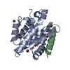
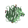
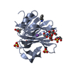
| Title | Crystal structure of a glycoside hydrolase in complex with cellobiose | |||||||||
 Components Components | Glycoside hydrolase family 45 protein | |||||||||
 Keywords Keywords |  HYDROLASE / substrate binding / cellulase / inhibitor HYDROLASE / substrate binding / cellulase / inhibitor | |||||||||
| Function / homology |  Function and homology informationcellulose binding / cellulase / cellulase activity / cellulose catabolic process / extracellular region Function and homology informationcellulose binding / cellulase / cellulase activity / cellulose catabolic process / extracellular regionSimilarity search - Function | |||||||||
| Biological species |  Thielavia terrestris NRRL 8126 (fungus) Thielavia terrestris NRRL 8126 (fungus) | |||||||||
| Method | X-RAY DIFFRACTION / SYNCHROTRON / MOLECULAR REPLACEMENT / Resolution: 1.36 Å | |||||||||
 Authors Authors | Gao, J. / Liu, W.D. / Zheng, Y.Y. / Chen, C.C. / Guo, R.T. | |||||||||
 Citation Citation | Journal: Enzyme Microb. Technol. / Year: 2017 Title: Characterization and crystal structure of a thermostable glycoside hydrolase family 45 1,4-beta-endoglucanase from Thielavia terrestris Authors: Gao, J. / Huang, J.W. / Li, Q. / Liu, W.D. / Ko, T.P. / Zheng, Y.Y. / Xiao, X. / Kuo, C.J. / Chen, C.C. / Guo, R.T. | |||||||||
| History |
|
- Structure visualization
| Structure viewer | Molecule: MolmilJmol/JSmol |
|---|
- Downloads & links
-Download
| PDBx/mmCIF format | 5gm9.cif.gz | 61.5 KB | Display | PDBx/mmCIF format |
|---|---|---|---|---|
| PDB format | pdb5gm9.ent.gz | 42.1 KB | Display | PDB format |
| PDBx/mmJSON format | 5gm9.json.gz | Tree view | PDBx/mmJSON format | |
| Others |  Other downloads Other downloads |
-Validation report
| Arichive directory | https://data.pdbj.org/pub/pdb/validation_reports/gm/5gm9ftp://data.pdbj.org/pub/pdb/validation_reports/gm/5gm9 | HTTPS FTP |
|---|
-Related structure data
| Related structure data |  5glxC  5glyC 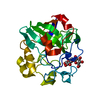 1oa7S C: citing same article ( S: Starting model for refinement |
|---|---|
| Similar structure data |


-Links
 PDBj
PDBj- Assembly
| Deposited unit | 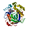
| ||||||||
|---|---|---|---|---|---|---|---|---|---|
| 1 |
| ||||||||
| Unit cell |
|
-Components
| #1: Protein | Mass: 22318.486 Da / Num. of mol.: 1 / Fragment: UNP residues 22-234 Source method: isolated from a genetically manipulated source Source: (gene. exp.) Thielavia terrestris NRRL 8126 (fungus)Strain: NRRL 8126 / Gene: THITE_2110957 / Production host: Komagataella pastoris (fungus) / Strain (production host): X33 / References: UniProt: G2QVH7 |
|---|---|
| #2: Polysaccharide | beta-D-glucopyranose-(1-4)-alpha-D-glucopyranose / alpha-cellobiose  , Oligosaccharide / Class: Metabolism / Mass: 342.297 Da / Num. of mol.: 1 , Oligosaccharide / Class: Metabolism / Mass: 342.297 Da / Num. of mol.: 1Source method: isolated from a genetically manipulated source Details: oligosaccharide / References: alpha-cellobiose |
| #3: Polysaccharide | beta-D-glucopyranose-(1-4)-beta-D-glucopyranose / beta-cellobiose  , Oligosaccharide / Class: Metabolism / Mass: 342.297 Da / Num. of mol.: 1 , Oligosaccharide / Class: Metabolism / Mass: 342.297 Da / Num. of mol.: 1Source method: isolated from a genetically manipulated source Details: oligosaccharide / References: beta-cellobiose |
| #4: Water | ChemComp-HOH / Water Mass: 18.015 Da / Num. of mol.: 237 / Source method: isolated from a natural source / Formula: H2O Mass: 18.015 Da / Num. of mol.: 237 / Source method: isolated from a natural source / Formula: H2O |
-Experimental details
-Experiment
| Experiment | Method: X-RAY DIFFRACTION / Number of used crystals: 1 |
|---|
- Sample preparation
| Crystal | Density Matthews: 2.39 Å3/Da / Density % sol: 48.58 % / Mosaicity: 0.329 ° |
|---|---|
| Crystal grow | Temperature: 298 K / Method: vapor diffusion, sitting drop / pH: 7.5 / Details: MgCl2, Tris-Cl, PEG4000 |
-Data collection
| Diffraction | Mean temperature: 100 K | ||||||||||||||||||||||||||||||||||||||||||||||||||||||||||||||||||
|---|---|---|---|---|---|---|---|---|---|---|---|---|---|---|---|---|---|---|---|---|---|---|---|---|---|---|---|---|---|---|---|---|---|---|---|---|---|---|---|---|---|---|---|---|---|---|---|---|---|---|---|---|---|---|---|---|---|---|---|---|---|---|---|---|---|---|---|
| Diffraction source | Source: SYNCHROTRON / Site: NSRRC  / Beamline: BL15A1 / Wavelength: 1 Å / Beamline: BL15A1 / Wavelength: 1 Å | ||||||||||||||||||||||||||||||||||||||||||||||||||||||||||||||||||
| Detector | Type: RAYONIX MX300HE / Detector: CCD / Date: Mar 16, 2016 | ||||||||||||||||||||||||||||||||||||||||||||||||||||||||||||||||||
| Radiation | Monochromator: GRAPHITE / Protocol: SINGLE WAVELENGTH / Monochromatic (M) / Laue (L): M / Scattering type: x-ray | ||||||||||||||||||||||||||||||||||||||||||||||||||||||||||||||||||
| Radiation wavelength | Wavelength: 1 Å / Relative weight: 1 | ||||||||||||||||||||||||||||||||||||||||||||||||||||||||||||||||||
| Reflection | Resolution: 1.36→25 Å / Num. obs: 46675 / % possible obs: 99.9 % / Redundancy: 10.8 % / Rmerge(I) obs: 0.035 / Net I/av σ(I): 57.314 / Net I/σ(I): 27.2 | ||||||||||||||||||||||||||||||||||||||||||||||||||||||||||||||||||
| Reflection shell |
|
- Processing
| Software |
| |||||||||||||||||||||||||||||||||||||||||||||||||||||||||||||||||||||||||||
|---|---|---|---|---|---|---|---|---|---|---|---|---|---|---|---|---|---|---|---|---|---|---|---|---|---|---|---|---|---|---|---|---|---|---|---|---|---|---|---|---|---|---|---|---|---|---|---|---|---|---|---|---|---|---|---|---|---|---|---|---|---|---|---|---|---|---|---|---|---|---|---|---|---|---|---|---|
| Refinement | Method to determine structure: MOLECULAR REPLACEMENT Starting model: 1OA7 Resolution: 1.36→25 Å / Cor.coef. Fo:Fc: 0.975 / Cor.coef. Fo:Fc free: 0.967 / SU B: 0.587 / SU ML: 0.025 / Cross valid method: THROUGHOUT / σ(F): 0 / ESU R: 0.044 / ESU R Free: 0.046 Details: HYDROGENS HAVE BEEN ADDED IN THE RIDING POSITIONS U VALUES : REFINED INDIVIDUALLY
| |||||||||||||||||||||||||||||||||||||||||||||||||||||||||||||||||||||||||||
| Solvent computation | Ion probe radii: 0.8 Å / Shrinkage radii: 0.8 Å / VDW probe radii: 1.2 Å | |||||||||||||||||||||||||||||||||||||||||||||||||||||||||||||||||||||||||||
| Displacement parameters | Biso max: 55.75 Å2 / Biso mean: 15.275 Å2 / Biso min: 8.07 Å2
| |||||||||||||||||||||||||||||||||||||||||||||||||||||||||||||||||||||||||||
| Refinement step | Cycle: final / Resolution: 1.36→25 Å
| |||||||||||||||||||||||||||||||||||||||||||||||||||||||||||||||||||||||||||
| Refine LS restraints |
| |||||||||||||||||||||||||||||||||||||||||||||||||||||||||||||||||||||||||||
| LS refinement shell | Resolution: 1.36→1.395 Å / Total num. of bins used: 20
|
