 Movie
Movie Controller


+ Open data

- Basic information
| Entry | Database: PDB / ID: 5fwg | ||||||
|---|---|---|---|---|---|---|---|
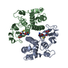
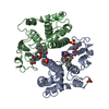
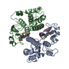
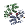
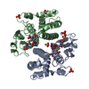
| Title | TETRA-(5-FLUOROTRYPTOPHANYL)-GLUTATHIONE TRANSFERASE | ||||||
 Components Components | TETRA-(5-FLUOROTRYPTOPHANYL)-GLUTATHIONE TRANSFERASE MU CLASS | ||||||
 Keywords Keywords |  TRANSFERASE / GLUTATHIONE TRANSFERASE / UNNATURAL AMINO ACID / 5-FLUOROTRYPTOPHAN / THREE-DIMENSIONAL STRUCTURE TRANSFERASE / GLUTATHIONE TRANSFERASE / UNNATURAL AMINO ACID / 5-FLUOROTRYPTOPHAN / THREE-DIMENSIONAL STRUCTURE | ||||||
| Function / homology |  Function and homology informationGlutathione conjugation / nitrobenzene metabolic process / cellular detoxification of nitrogen compound / hepoxilin biosynthetic process / glutathione binding / glutathione derivative biosynthetic process / response to metal ion / prostaglandin metabolic process / nickel cation binding / glutathione transferase ...Glutathione conjugation / nitrobenzene metabolic process / cellular detoxification of nitrogen compound / hepoxilin biosynthetic process / glutathione binding / glutathione derivative biosynthetic process / response to metal ion / prostaglandin metabolic process / nickel cation binding / glutathione transferase / glutathione transferase activity / response to axon injury / response to amino acid / xenobiotic catabolic process / glutathione metabolic process / steroid binding / response to lead ion / cellular response to xenobiotic stimulus / sensory perception of smell / response to ethanol / response to xenobiotic stimulus / protein kinase binding / enzyme binding / protein homodimerization activity / protein-containing complex / extracellular region / identical protein binding / cytosol / cytoplasm Function and homology informationGlutathione conjugation / nitrobenzene metabolic process / cellular detoxification of nitrogen compound / hepoxilin biosynthetic process / glutathione binding / glutathione derivative biosynthetic process / response to metal ion / prostaglandin metabolic process / nickel cation binding / glutathione transferase ...Glutathione conjugation / nitrobenzene metabolic process / cellular detoxification of nitrogen compound / hepoxilin biosynthetic process / glutathione binding / glutathione derivative biosynthetic process / response to metal ion / prostaglandin metabolic process / nickel cation binding / glutathione transferase / glutathione transferase activity / response to axon injury / response to amino acid / xenobiotic catabolic process / glutathione metabolic process / steroid binding / response to lead ion / cellular response to xenobiotic stimulus / sensory perception of smell / response to ethanol / response to xenobiotic stimulus / protein kinase binding / enzyme binding / protein homodimerization activity / protein-containing complex / extracellular region / identical protein binding / cytosol / cytoplasmSimilarity search - Function | ||||||
| Biological species |  Rattus norvegicus (Norway rat) Rattus norvegicus (Norway rat) | ||||||
| Method | X-RAY DIFFRACTION / MOLECULAR MODIFICATION / Resolution: 2 Å | ||||||
 Authors Authors | Parsons, J.F. / Xiao, G. / Armstrong, R.N. / Gilliland, G.L. | ||||||
 Citation Citation | Journal: Biochemistry / Year: 1998 Title: Enzymes harboring unnatural amino acids: mechanistic and structural analysis of the enhanced catalytic activity of a glutathione transferase containing 5-fluorotryptophan. Authors: Parsons, J.F. / Xiao, G. / Gilliland, G.L. / Armstrong, R.N. #1: Journal: J.Biol.Chem. / Year: 1993Title: Tyrosine 115 Participates Both in Chemical and Physical Steps of the Catalytic Mechanism of a Glutathione S-Transferase Authors: Johnson, W.W. / Liu, S. / Ji, X. / Gilliland, G.L. / Armstrong, R.N. #2: Journal: J.Biol.Chem. / Year: 1992Title: Contribution of Tyrosine 6 to the Catalytic Mechanism of Isoenzyme 3-3 of Glutathione S-Transferase Authors: Liu, S. / Zhang, P. / Ji, X. / Johnson, W.W. / Gilliland, G.L. / Armstrong, R.N. #3: Journal: Biochemistry / Year: 1992Title: The Three-Dimensional Structure of a Glutathione S-Transferase from the Mu Gene Class. Structural Analysis of the Binary Complex of Isoenzyme 3-3 and Glutathione at 2.2-A Resolution Authors: Ji, X. / Zhang, P. / Armstrong, R.N. / Gilliland, G.L. | ||||||
| History |
|
- Structure visualization
| Structure viewer | Molecule: MolmilJmol/JSmol |
|---|
- Downloads & links
-Download
| PDBx/mmCIF format | 5fwg.cif.gz | 110.2 KB | Display | PDBx/mmCIF format |
|---|---|---|---|---|
| PDB format | pdb5fwg.ent.gz | 88.9 KB | Display | PDB format |
| PDBx/mmJSON format | 5fwg.json.gz | Tree view | PDBx/mmJSON format | |
| Others |  Other downloads Other downloads |
-Validation report
| Arichive directory | https://data.pdbj.org/pub/pdb/validation_reports/fw/5fwgftp://data.pdbj.org/pub/pdb/validation_reports/fw/5fwg | HTTPS FTP |
|---|
-Related structure data
| Related structure data |  3gstS S: Starting model for refinement |
|---|---|
| Similar structure data |


-Links
 PDBj
PDBj

- Assembly
| Deposited unit | 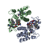
| ||||||||
|---|---|---|---|---|---|---|---|---|---|
| 1 |
| ||||||||
| Unit cell |
| ||||||||
| Noncrystallographic symmetry (NCS) | NCS oper: (Code: given Matrix: (0.841869, -0.090312, 0.532071), Vector : |
-Components
| #1: Protein | Mass: 25890.756 Da / Num. of mol.: 2 / Mutation: 4 TRYPTOPHANS MUTATED TO 5-FLUOROTRYPTOPHANS Source method: isolated from a genetically manipulated source Source: (gene. exp.) Rattus norvegicus (Norway rat) / Cell line: BL21 / Gene: CDNA INSERT OF 3-3 (M1-1) ENZY / Organ: LIVER / Plasmid: PSW1GST33 / Species (production host): Escherichia coli / Gene (production host): CDNA INSERT OF 3-3 (M1-1) ENZYME / Production host:  Escherichia coli BL21(DE3) (bacteria) / Strain (production host): BL21 (DE3) / References: UniProt: P04905, glutathione transferase Escherichia coli BL21(DE3) (bacteria) / Strain (production host): BL21 (DE3) / References: UniProt: P04905, glutathione transferase#2: Chemical |   Mass: 501.552 Da / Num. of mol.: 2 / Source method: obtained synthetically / Formula: C24H27N3O7S Mass: 501.552 Da / Num. of mol.: 2 / Source method: obtained synthetically / Formula: C24H27N3O7S#3: Water | ChemComp-HOH / | Water Mass: 18.015 Da / Num. of mol.: 371 / Source method: isolated from a natural source / Formula: H2O Mass: 18.015 Da / Num. of mol.: 371 / Source method: isolated from a natural source / Formula: H2O |
|---|
-Experimental details
-Experiment
| Experiment | Method: X-RAY DIFFRACTION / Number of used crystals: 1 |
|---|
- Sample preparation
| Crystal | Density Matthews: 2.31 Å3/Da / Density % sol: 47 % | |||||||||||||||||||||||||||||||||||
|---|---|---|---|---|---|---|---|---|---|---|---|---|---|---|---|---|---|---|---|---|---|---|---|---|---|---|---|---|---|---|---|---|---|---|---|---|
| Crystal grow | pH: 7.6 / Details: pH 7.6 | |||||||||||||||||||||||||||||||||||
| Crystal grow | *PLUS Temperature: 22 ℃ / Method: vapor diffusion, hanging drop | |||||||||||||||||||||||||||||||||||
| Components of the solutions | *PLUS
|
-Data collection
| Diffraction | Mean temperature: 103 K |
|---|---|
| Diffraction source | Source: ROTATING ANODE / Type: RIGAKU RUH2R / Wavelength: 1.5418 |
| Detector | Type: RIGAKU / Detector: IMAGE PLATE / Date: Apr 8, 1997 / Details: YALE MIRRORS |
| Radiation | Monochromator: YALE MIRRORS / Monochromatic (M) / Laue (L): M / Scattering type: x-ray |
| Radiation wavelength | Wavelength: 1.5418 Å / Relative weight: 1 |
| Reflection | Resolution: 2→65 Å / Num. obs: 184888 / % possible obs: 94.9 % / Observed criterion σ(I): 5 / Redundancy: 6.24 % / Biso Wilson estimate: 27.8 Å2 / Rmerge(I) obs: 0.066 / Net I/σ(I): 20 |
| Reflection shell | Resolution: 2→2.07 Å / Redundancy: 2 % / Rmerge(I) obs: 0.137 / Mean I/σ(I) obs: 15 / % possible all: 68.3 |
| Reflection | *PLUS Num. obs: 29587 / % possible obs: 95 % / Num. measured all: 184888 |
| Reflection shell | *PLUS % possible obs: 68.3 % / Mean I/σ(I) obs: 9.1 |
- Processing
| Software |
| ||||||||||||||||||||||||||||||||||||||||||||||||||
|---|---|---|---|---|---|---|---|---|---|---|---|---|---|---|---|---|---|---|---|---|---|---|---|---|---|---|---|---|---|---|---|---|---|---|---|---|---|---|---|---|---|---|---|---|---|---|---|---|---|---|---|
| Refinement | Method to determine structure: MOLECULAR MODIFICATION Starting model: PDB ENTRY 3GST Resolution: 2→65 Å / Isotropic thermal model: TNT BCORREL V1.0 / σ(F): 2 / Stereochemistry target values: TNT PROTGEO /
| ||||||||||||||||||||||||||||||||||||||||||||||||||
| Solvent computation | Solvent model: BABINET SCALING / Bsol: 71.4 Å2 / ksol: 0.814 e/Å3 | ||||||||||||||||||||||||||||||||||||||||||||||||||
| Refinement step | Cycle: LAST / Resolution: 2→65 Å
| ||||||||||||||||||||||||||||||||||||||||||||||||||
| Refine LS restraints |
| ||||||||||||||||||||||||||||||||||||||||||||||||||
| Software | *PLUS Name: TNT / Version: 5E / Classification: refinement | ||||||||||||||||||||||||||||||||||||||||||||||||||
| Refinement | *PLUS Num. reflection obs: 28444 / Rfactor obs: 0.181 | ||||||||||||||||||||||||||||||||||||||||||||||||||
| Solvent computation | *PLUS | ||||||||||||||||||||||||||||||||||||||||||||||||||
| Displacement parameters | *PLUS | ||||||||||||||||||||||||||||||||||||||||||||||||||
| Refine LS restraints | *PLUS
|
