 Movie
Movie Controller

[English] 日本語
 Yorodumi
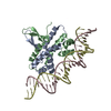
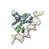
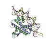
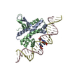
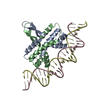
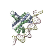
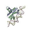
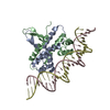
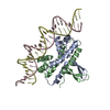
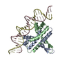
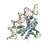
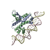
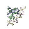
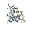
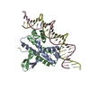
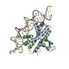
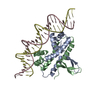
- PDB-5e3m: Crystal structure of Fis bound to 27bp DNA F35 (AAATTAGTTTGAATCTC... -
+ Open data

- Basic information
| Entry | Database: PDB / ID: 5e3m | ||||||
|---|---|---|---|---|---|---|---|
| Title | Crystal structure of Fis bound to 27bp DNA F35 (AAATTAGTTTGAATCTCGAGCTAATTT) | ||||||
 Components Components |
| ||||||
 Keywords Keywords | DNA BINDING PROTEIN/DNA /  Protein-DNA complex / HTH domain / minor groove compression / DNA bending / indirect recognition / DNA BINDING PROTEIN-DNA complex Protein-DNA complex / HTH domain / minor groove compression / DNA bending / indirect recognition / DNA BINDING PROTEIN-DNA complex | ||||||
| Function / homology |  Function and homology information Function and homology informationinvertasome / positive regulation of DNA recombination / sequence-specific DNA binding, bending / provirus excision / nucleoid / DNA-binding transcription repressor activity / DNA-binding transcription activator activity / chromosome organization / core promoter sequence-specific DNA binding / protein-DNA complex ...invertasome / positive regulation of DNA recombination / sequence-specific DNA binding, bending / provirus excision / nucleoid / DNA-binding transcription repressor activity / DNA-binding transcription activator activity / chromosome organization / core promoter sequence-specific DNA binding / protein-DNA complex / response to radiation / nucleosome / sequence-specific DNA binding / transcription cis-regulatory region binding / DNA-templated transcription / regulation of DNA-templated transcription / protein homodimerization activity / DNA binding / cytosolSimilarity search - Function | ||||||
| Biological species |  Escherichia coli (E. coli) Escherichia coli (E. coli)synthetic construct (others) | ||||||
| Method | X-RAY DIFFRACTION / SYNCHROTRON / MOLECULAR REPLACEMENT / molecular replacement / Resolution: 2.886 Å | ||||||
 Authors Authors | Stella, S. / Hancock, S.P. / Cascio, D. / Johnson, R.C. | ||||||
| Funding support |  United States, 1items United States, 1items
| ||||||
 Citation Citation | Journal: Plos One / Year: 2016 Title: DNA Sequence Determinants Controlling Affinity, Stability and Shape of DNA Complexes Bound by the Nucleoid Protein Fis. Authors: Hancock, S.P. / Stella, S. / Cascio, D. / Johnson, R.C. | ||||||
| History |
|
- Structure visualization
| Structure viewer | Molecule: MolmilJmol/JSmol |
|---|
- Downloads & links
-Download
| PDBx/mmCIF format | 5e3m.cif.gz | 140.6 KB | Display | PDBx/mmCIF format |
|---|---|---|---|---|
| PDB format | pdb5e3m.ent.gz | 113.8 KB | Display | PDB format |
| PDBx/mmJSON format | 5e3m.json.gz | Tree view | PDBx/mmJSON format | |
| Others |  Other downloads Other downloads |
-Validation report
| Arichive directory | https://data.pdbj.org/pub/pdb/validation_reports/e3/5e3mftp://data.pdbj.org/pub/pdb/validation_reports/e3/5e3m | HTTPS FTP |
|---|
-Related structure data


-Links
 PDBj
PDBj


- Assembly
| Deposited unit | 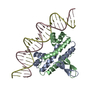
| ||||||||
|---|---|---|---|---|---|---|---|---|---|
| 1 |
| ||||||||
| Unit cell |
|
-Components
| #1: Protein | Mass: 11252.918 Da / Num. of mol.: 2 Source method: isolated from a genetically manipulated source Source: (gene. exp.) Escherichia coli (E. coli) / Strain: K12 / Gene: fis, b3261, JW3229 / Plasmid: pET11a / Production host: Escherichia coli (E. coli) / Strain (production host): BL21(DE3) / References: UniProt: P0A6R3#2: DNA chain | | Mass: 8304.393 Da / Num. of mol.: 1 / Source method: obtained synthetically / Source: (synth.) synthetic construct (others) #3: DNA chain | | Mass: 8282.397 Da / Num. of mol.: 1 / Source method: obtained synthetically / Source: (synth.) synthetic construct (others) #4: Water | ChemComp-HOH / | Water Mass: 18.015 Da / Num. of mol.: 7 / Source method: isolated from a natural source / Formula: H2O Mass: 18.015 Da / Num. of mol.: 7 / Source method: isolated from a natural source / Formula: H2O |
|---|
-Experimental details
-Experiment
| Experiment | Method: X-RAY DIFFRACTION / Number of used crystals: 1 |
|---|
- Sample preparation
| Crystal | Density Matthews: 4.06 Å3/Da / Density % sol: 69.68 % |
|---|---|
| Crystal grow | Temperature: 277 K / Method: vapor diffusion, hanging drop / pH: 8.5 Details: 0.2 M Sodium citrate, 0.1 M TRIS-HCl pH 8.5, 36% PEG 400 |
-Data collection
| Diffraction | Mean temperature: 100 K | |||||||||||||||||||||||||||
|---|---|---|---|---|---|---|---|---|---|---|---|---|---|---|---|---|---|---|---|---|---|---|---|---|---|---|---|---|
| Diffraction source | Source: SYNCHROTRON / Site: ALS / Beamline: 8.2.2 / Wavelength: 1 Å | |||||||||||||||||||||||||||
| Detector | Type: ADSC QUANTUM 315 / Detector: CCD / Date: Mar 5, 2007 | |||||||||||||||||||||||||||
| Radiation | Monochromator: Si (111) / Protocol: SINGLE WAVELENGTH / Monochromatic (M) / Laue (L): M / Scattering type: x-ray | |||||||||||||||||||||||||||
| Radiation wavelength | Wavelength: 1 Å / Relative weight: 1 | |||||||||||||||||||||||||||
| Reflection | Resolution: 2.88→40.412 Å / Num. obs: 13581 / % possible obs: 94.4 % / Redundancy: 6.6 % / Biso Wilson estimate: 35.76 Å2 / CC1/2: 0.998 / Rmerge(I) obs: 0.082 / Rpim(I) all: 0.034 / Net I/σ(I): 13.7 / Num. measured all: 89910 | |||||||||||||||||||||||||||
| Reflection shell | Diffraction-ID: 1 / Rejects: 0
|
-Phasing
| Phasing | Method: molecular replacement |
|---|
- Processing
| Software |
| ||||||||||||||||||||||||||||||||||||||||||
|---|---|---|---|---|---|---|---|---|---|---|---|---|---|---|---|---|---|---|---|---|---|---|---|---|---|---|---|---|---|---|---|---|---|---|---|---|---|---|---|---|---|---|---|
| Refinement | Method to determine structure: MOLECULAR REPLACEMENT / Resolution: 2.886→40.412 Å / SU ML: 0.35 / Cross valid method: FREE R-VALUE / σ(F): 1.34 / Phase error: 20.91 / Stereochemistry target values: ML
| ||||||||||||||||||||||||||||||||||||||||||
| Solvent computation | Shrinkage radii: 0.9 Å / VDW probe radii: 1.11 Å / Solvent model: FLAT BULK SOLVENT MODEL | ||||||||||||||||||||||||||||||||||||||||||
| Displacement parameters | Biso max: 163.29 Å2 / Biso mean: 43.4773 Å2 / Biso min: 8.38 Å2 | ||||||||||||||||||||||||||||||||||||||||||
| Refinement step | Cycle: final / Resolution: 2.886→40.412 Å
| ||||||||||||||||||||||||||||||||||||||||||
| Refine LS restraints |
| ||||||||||||||||||||||||||||||||||||||||||
| LS refinement shell | Refine-ID: X-RAY DIFFRACTION / Total num. of bins used: 5
| ||||||||||||||||||||||||||||||||||||||||||
| Refinement TLS params. | Method: refined / Origin x: 32.5154 Å / Origin y: -10.9848 Å / Origin z: 20.7565 Å
| ||||||||||||||||||||||||||||||||||||||||||
| Refinement TLS group |
|
