 Movie
Movie Controller

[English] 日本語
 Yorodumi
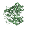
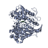
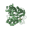
- PDB-5e3a: Structure of human DPP3 in complex with opioid peptide leu-enkephalin -
+ Open data

- Basic information
| Entry | Database: PDB / ID: 5e3a | ||||||||||||
|---|---|---|---|---|---|---|---|---|---|---|---|---|---|
| Title | Structure of human DPP3 in complex with opioid peptide leu-enkephalin | ||||||||||||
 Components Components |
| ||||||||||||
 Keywords Keywords |  HYDROLASE / Peptide-complex / Zinc-hydrolase / opioid-peptide / peptidase HYDROLASE / Peptide-complex / Zinc-hydrolase / opioid-peptide / peptidase | ||||||||||||
| Function / homology |  Function and homology information Function and homology informationsynaptic vesicle lumen / neuronal dense core vesicle lumen / dipeptidyl-peptidase III / chromaffin granule lumen / opioid receptor binding / opioid peptide activity / synaptic signaling via neuropeptide / general adaptation syndrome, behavioral process / aggressive behavior / positive regulation of behavioral fear response ...synaptic vesicle lumen / neuronal dense core vesicle lumen / dipeptidyl-peptidase III / chromaffin granule lumen / opioid receptor binding / opioid peptide activity / synaptic signaling via neuropeptide / general adaptation syndrome, behavioral process / aggressive behavior / positive regulation of behavioral fear response / G protein-coupled opioid receptor signaling pathway / symmetric synapse / cell body fiber / response to epinephrine / sensory perception / cellular response to vitamin D / neuropeptide hormone activity / metalloexopeptidase activity / dipeptidyl-peptidase activity / locomotory exploration behavior / transmission of nerve impulse / startle response / neuropeptide signaling pathway / behavioral fear response / glial cell proliferation / axon terminus / aminopeptidase activity / cellular response to cAMP / sensory perception of pain / cellular response to transforming growth factor beta stimulus / Peptide ligand-binding receptors / response to nicotine / Post-translational protein phosphorylation / protein catabolic process / response to toxic substance / osteoblast differentiation / cellular response to virus / response to calcium ion / Regulation of Insulin-like Growth Factor (IGF) transport and uptake by Insulin-like Growth Factor Binding Proteins (IGFBPs) / KEAP1-NFE2L2 pathway / response to estradiol / cellular response to oxidative stress / Neddylation / G alpha (i) signalling events / chemical synaptic transmission / perikaryon / response to ethanol / response to lipopolysaccharide / response to hypoxia / endoplasmic reticulum lumen / dendrite / neuronal cell body / signal transduction / proteolysis / extracellular exosome / zinc ion binding / extracellular region / plasma membrane / cytosol / cytoplasmSimilarity search - Function | ||||||||||||
| Biological species |  Homo sapiens (human) Homo sapiens (human) | ||||||||||||
| Method | X-RAY DIFFRACTION / SYNCHROTRON / MOLECULAR REPLACEMENT / Resolution: 2.05 Å | ||||||||||||
 Authors Authors | Kumar, P. / Reithofer, V. / Reisinger, M. / Pavkov-Keller, T. / Wallner, S. / Macheroux, P. / Gruber, K. | ||||||||||||
| Funding support |  Austria, 1items Austria, 1items
| ||||||||||||
 Citation Citation | Journal: Sci Rep / Year: 2016 Title: Substrate complexes of human dipeptidyl peptidase III reveal the mechanism of enzyme inhibition. Authors: Kumar, P. / Reithofer, V. / Reisinger, M. / Wallner, S. / Pavkov-Keller, T. / Macheroux, P. / Gruber, K. | ||||||||||||
| History |
|
- Structure visualization
| Structure viewer | Molecule: MolmilJmol/JSmol |
|---|
- Downloads & links
-Download
| PDBx/mmCIF format | 5e3a.cif.gz | 166.8 KB | Display | PDBx/mmCIF format |
|---|---|---|---|---|
| PDB format | pdb5e3a.ent.gz | 129.5 KB | Display | PDB format |
| PDBx/mmJSON format | 5e3a.json.gz | Tree view | PDBx/mmJSON format | |
| Others |  Other downloads Other downloads |
-Validation report
| Arichive directory | https://data.pdbj.org/pub/pdb/validation_reports/e3/5e3aftp://data.pdbj.org/pub/pdb/validation_reports/e3/5e3a | HTTPS FTP |
|---|
-Related structure data
| Related structure data | 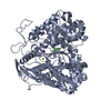 5e2qC  5e33C  5e3cC 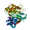 5egyC  5ehhC  3t6bS S: Starting model for refinement C: citing same article ( |
|---|---|
| Similar structure data |


-Links
 PDBj
PDBj


- Assembly
| Deposited unit | 
| |||||||||
|---|---|---|---|---|---|---|---|---|---|---|
| 1 |
| |||||||||
| Unit cell |
| |||||||||
| Components on special symmetry positions |
|
-Components
-Protein / Protein/peptide , 2 types, 2 molecules AB
| #1: Protein | DPP3 / Dipeptidyl aminopeptidase III / Dipeptidyl arylamidase III / Dipeptidyl peptidase III / DPP III / ...Dipeptidyl aminopeptidase III / Dipeptidyl arylamidase III / Dipeptidyl peptidase III / DPP III / Enkephalinase B Mass: 81548.688 Da / Num. of mol.: 1 / Mutation: C19S, E207C, E451A, S491C, C519S, C654S Source method: isolated from a genetically manipulated source Source: (gene. exp.) Homo sapiens (human) / Gene: DPP3 / Plasmid: PET28MHL / Production host:  Escherichia coli BL21(DE3) (bacteria) / References: UniProt: Q9NY33, dipeptidyl-peptidase III Escherichia coli BL21(DE3) (bacteria) / References: UniProt: Q9NY33, dipeptidyl-peptidase III |
|---|---|
| #2: Protein/peptide | Mass: 555.624 Da / Num. of mol.: 1 / Source method: obtained synthetically / Details: opioid peptide from human source / Source: (synth.) Homo sapiens (human) / References: UniProt: P01210*PLUS |
-Non-polymers , 4 types, 367 molecules 


| #3: Chemical | ChemComp-ZN /  Mass: 65.409 Da / Num. of mol.: 1 / Source method: obtained synthetically / Formula: Zn Mass: 65.409 Da / Num. of mol.: 1 / Source method: obtained synthetically / Formula: Zn | ||||
|---|---|---|---|---|---|
| #4: Chemical |  Mass: 24.305 Da / Num. of mol.: 2 / Source method: obtained synthetically / Formula: Mg Mass: 24.305 Da / Num. of mol.: 2 / Source method: obtained synthetically / Formula: Mg#5: Chemical | ChemComp-K / |  Mass: 39.098 Da / Num. of mol.: 1 / Source method: obtained synthetically / Formula: K Mass: 39.098 Da / Num. of mol.: 1 / Source method: obtained synthetically / Formula: K#6: Water | ChemComp-HOH / | WaterMass: 18.015 Da / Num. of mol.: 363 / Source method: isolated from a natural source / Formula: H2O |
-Experimental details
-Experiment
| Experiment | Method: X-RAY DIFFRACTION |
|---|
- Sample preparation
| Crystal | Density Matthews: 2.5 Å3/Da / Density % sol: 50.82 % |
|---|---|
| Crystal grow | Temperature: 293 K / Method: vapor diffusion, sitting drop / pH: 8.2 Details: 0.056M SODIUM PHOSPHATE MONOBASIC MONOHYDRATE, 1.344M POTASSIUM PHOSPHATE DIBASIC PH range: 8.2 |
-Data collection
| Diffraction | Mean temperature: 100 K |
|---|---|
| Diffraction source | Source: SYNCHROTRON / Site: ESRF  / Beamline: ID29 / Wavelength: 0.97239 Å / Beamline: ID29 / Wavelength: 0.97239 Å |
| Detector | Type: DECTRIS PILATUS 6M / Detector: PIXEL / Date: Apr 20, 2015 |
| Radiation | Monochromator: liquid nitrogen cooled channel-cut silicon monochromator Protocol: SINGLE WAVELENGTH / Monochromatic (M) / Laue (L): M / Scattering type: x-ray |
| Radiation wavelength | Wavelength: 0.97239 Å / Relative weight: 1 |
| Reflection | Resolution: 2.05→39.58 Å / Num. all: 48267 / Num. obs: 48267 / % possible obs: 95.09 % / Redundancy: 3 % / Biso Wilson estimate: 32.96 Å2 / Rmerge(I) obs: 0.08875 / Rsym value: 0.08875 / Net I/σ(I): 10.73 |
| Reflection shell | Resolution: 2.05→2.121 Å / Redundancy: 3.1 % / Rmerge(I) obs: 0.6197 / Mean I/σ(I) obs: 1.85 / % possible all: 95.45 |
- Processing
| Software |
| ||||||||||||||||||||||||||||||||||||||||||||||||||||||||||||||||||||||||||||||||||||||||||||||||||||||||||||||||||||||||||||||
|---|---|---|---|---|---|---|---|---|---|---|---|---|---|---|---|---|---|---|---|---|---|---|---|---|---|---|---|---|---|---|---|---|---|---|---|---|---|---|---|---|---|---|---|---|---|---|---|---|---|---|---|---|---|---|---|---|---|---|---|---|---|---|---|---|---|---|---|---|---|---|---|---|---|---|---|---|---|---|---|---|---|---|---|---|---|---|---|---|---|---|---|---|---|---|---|---|---|---|---|---|---|---|---|---|---|---|---|---|---|---|---|---|---|---|---|---|---|---|---|---|---|---|---|---|---|---|---|
| Refinement | Method to determine structure: MOLECULAR REPLACEMENT Starting model: 3T6B Resolution: 2.05→39.576 Å / SU ML: 0.33 / Cross valid method: FREE R-VALUE / σ(F): 1.35 / ESU R: 0.2088 / ESU R Free: 0.259 / Phase error: 30.47 / Stereochemistry target values: ML
| ||||||||||||||||||||||||||||||||||||||||||||||||||||||||||||||||||||||||||||||||||||||||||||||||||||||||||||||||||||||||||||||
| Solvent computation | Shrinkage radii: 0.9 Å / VDW probe radii: 1.11 Å / Solvent model: FLAT BULK SOLVENT MODEL | ||||||||||||||||||||||||||||||||||||||||||||||||||||||||||||||||||||||||||||||||||||||||||||||||||||||||||||||||||||||||||||||
| Displacement parameters | Biso mean: 37.2 Å2 | ||||||||||||||||||||||||||||||||||||||||||||||||||||||||||||||||||||||||||||||||||||||||||||||||||||||||||||||||||||||||||||||
| Refinement step | Cycle: LAST / Resolution: 2.05→39.576 Å
| ||||||||||||||||||||||||||||||||||||||||||||||||||||||||||||||||||||||||||||||||||||||||||||||||||||||||||||||||||||||||||||||
| Refine LS restraints |
| ||||||||||||||||||||||||||||||||||||||||||||||||||||||||||||||||||||||||||||||||||||||||||||||||||||||||||||||||||||||||||||||
| LS refinement shell |
|
