 Movie
Movie Controller

[English] 日本語
 Yorodumi
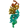
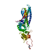
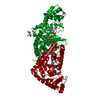
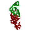
- PDB-5e26: Crystal structure of human PANK2: the catalytic core domain in co... -
+ Open data

- Basic information
| Entry | Database: PDB / ID: 5.0E+26 | ||||||
|---|---|---|---|---|---|---|---|
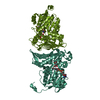
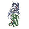
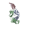
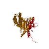
| Title | Crystal structure of human PANK2: the catalytic core domain in complex with pantothenate and adenosine diphosphate | ||||||
 Components Components | Pantothenate kinase 2, mitochondrial | ||||||
 Keywords Keywords | TRANSFERASE / PANK2 / Structural Genomics / Structural Genomics Consortium / SGC | ||||||
| Function / homology |  Function and homology information Function and homology informationpantothenate metabolic process / regulation of triglyceride metabolic process / Coenzyme A biosynthesis / regulation of bile acid metabolic process / : / pantothenate kinase / pantothenate kinase activity / regulation of fatty acid metabolic process / coenzyme A biosynthetic process / spermatid development ...pantothenate metabolic process / regulation of triglyceride metabolic process / Coenzyme A biosynthesis / regulation of bile acid metabolic process / : / pantothenate kinase / pantothenate kinase activity / regulation of fatty acid metabolic process / coenzyme A biosynthetic process / spermatid development / aerobic respiration / regulation of mitochondrial membrane potential / mitochondrial intermembrane space / angiogenesis / phosphorylation / mitochondrion / ATP binding / nucleus / cytosolSimilarity search - Function | ||||||
| Biological species |  Homo sapiens (human) Homo sapiens (human) | ||||||
| Method | X-RAY DIFFRACTION / SYNCHROTRON / MOLECULAR REPLACEMENT / molecular replacement / Resolution: 2.14 Å | ||||||
 Authors Authors | DONG, A. / LOPPNAU, P. / RAVICHANDRAN, M. / CHENG, C. / TEMPEL, W. / SEITOVA, A. / HUTCHINSON, A. / HONG, B.S. / Bountra, C. / Arrowsmith, C.H. ...DONG, A. / LOPPNAU, P. / RAVICHANDRAN, M. / CHENG, C. / TEMPEL, W. / SEITOVA, A. / HUTCHINSON, A. / HONG, B.S. / Bountra, C. / Arrowsmith, C.H. / Edwards, A.M. / BROWN, P.J. / Structural Genomics Consortium (SGC) | ||||||
 Citation Citation | Journal: to be published Title: Crystal structure of human PANK2: the catalytic core domain in complex with pantothenate and adenosine diphosphate Authors: LOPPNAU, P. / DONG, A. / RAVICHANDRAN, M. / CHENG, C. / TEMPEL, W. / SEITOVA, A. / HUTCHINSON, A. / HONG, B.S. / Bountra, C. / Arrowsmith, C.H. / Edwards, A.M. / BROWN, P.J. / Structural ...Authors: LOPPNAU, P. / DONG, A. / RAVICHANDRAN, M. / CHENG, C. / TEMPEL, W. / SEITOVA, A. / HUTCHINSON, A. / HONG, B.S. / Bountra, C. / Arrowsmith, C.H. / Edwards, A.M. / BROWN, P.J. / Structural Genomics Consortium (SGC) | ||||||
| History |
|
- Structure visualization
| Structure viewer | Molecule: MolmilJmol/JSmol |
|---|
- Downloads & links
-Download
| PDBx/mmCIF format | 5e26.cif.gz | 295.5 KB | Display | PDBx/mmCIF format |
|---|---|---|---|---|
| PDB format | pdb5e26.ent.gz | 236.5 KB | Display | PDB format |
| PDBx/mmJSON format | 5e26.json.gz | Tree view | PDBx/mmJSON format | |
| Others |  Other downloads Other downloads |
-Validation report
| Arichive directory | https://data.pdbj.org/pub/pdb/validation_reports/e2/5e26ftp://data.pdbj.org/pub/pdb/validation_reports/e2/5e26 | HTTPS FTP |
|---|
-Related structure data
| Related structure data |  3smsS S: Starting model for refinement |
|---|---|
| Similar structure data |


-Links
 PDBj
PDBj- Assembly
| Deposited unit | 
| ||||||||
|---|---|---|---|---|---|---|---|---|---|
| 1 | 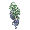
| ||||||||
| 2 | 
| ||||||||
| Unit cell |
|
-Components
-Protein , 1 types, 4 molecules ABCD
| #1: Protein | / hPanK2 / Pantothenic acid kinase 2 Mass: 40692.688 Da / Num. of mol.: 4 / Fragment: UNP residues 205-568 Source method: isolated from a genetically manipulated source Source: (gene. exp.) Homo sapiens (human) / Gene: PANK2, C20orf48 / Plasmid: pFBOH-MHL / Cell line (production host): sp9 / Production host:   Spodoptera frugiperda (fall armyworm) / Strain (production host): BVES / References: UniProt: Q9BZ23, pantothenate kinase Spodoptera frugiperda (fall armyworm) / Strain (production host): BVES / References: UniProt: Q9BZ23, pantothenate kinase |
|---|
-Non-polymers , 6 types, 288 molecules 
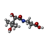


| #2: Chemical | ChemComp-ADP / Adenosine diphosphate Mass: 427.201 Da / Num. of mol.: 4 / Source method: obtained synthetically / Formula: C10H15N5O10P2 / Comment: ADP, energy-carrying molecule*YM Mass: 427.201 Da / Num. of mol.: 4 / Source method: obtained synthetically / Formula: C10H15N5O10P2 / Comment: ADP, energy-carrying molecule*YM#3: Chemical | ChemComp-PAU / Pantothenic acid Type: peptide-like / Mass: 219.235 Da / Num. of mol.: 4 / Source method: obtained synthetically / Formula: C9H17NO5 Type: peptide-like / Mass: 219.235 Da / Num. of mol.: 4 / Source method: obtained synthetically / Formula: C9H17NO5#4: Chemical | Glycerol Mass: 92.094 Da / Num. of mol.: 2 / Source method: obtained synthetically / Formula: C3H8O3 Mass: 92.094 Da / Num. of mol.: 2 / Source method: obtained synthetically / Formula: C3H8O3#5: Chemical | ChemComp-UNX /  Mass: 35.453 Da / Num. of mol.: 9 / Source method: obtained synthetically Mass: 35.453 Da / Num. of mol.: 9 / Source method: obtained synthetically#6: Chemical | Chloride Mass: 35.453 Da / Num. of mol.: 2 / Source method: obtained synthetically / Formula: Cl Mass: 35.453 Da / Num. of mol.: 2 / Source method: obtained synthetically / Formula: Cl#7: Water | ChemComp-HOH / | WaterMass: 18.015 Da / Num. of mol.: 267 / Source method: isolated from a natural source / Formula: H2O |
|---|
-Experimental details
-Experiment
| Experiment | Method: X-RAY DIFFRACTION / Number of used crystals: 1 |
|---|
- Sample preparation
| Crystal | Density Matthews: 2.62 Å3/Da / Density % sol: 52.98 % |
|---|---|
| Crystal grow | Temperature: 291 K / Method: vapor diffusion, sitting drop / pH: 6.5 / Details: 2.4M Na Malonate pH 6.5, 0.1M TCEP HCl |
-Data collection
| Diffraction | Mean temperature: 100 K | |||||||||||||||||||||||||||||||||||||||||||||||||||||||||||||||||||||||||||||||||||||||||||||||||||||||||||||||||||||||||||||||||||||||||||||||||||||||||||||||||||||||||||||||||||||||||||||
|---|---|---|---|---|---|---|---|---|---|---|---|---|---|---|---|---|---|---|---|---|---|---|---|---|---|---|---|---|---|---|---|---|---|---|---|---|---|---|---|---|---|---|---|---|---|---|---|---|---|---|---|---|---|---|---|---|---|---|---|---|---|---|---|---|---|---|---|---|---|---|---|---|---|---|---|---|---|---|---|---|---|---|---|---|---|---|---|---|---|---|---|---|---|---|---|---|---|---|---|---|---|---|---|---|---|---|---|---|---|---|---|---|---|---|---|---|---|---|---|---|---|---|---|---|---|---|---|---|---|---|---|---|---|---|---|---|---|---|---|---|---|---|---|---|---|---|---|---|---|---|---|---|---|---|---|---|---|---|---|---|---|---|---|---|---|---|---|---|---|---|---|---|---|---|---|---|---|---|---|---|---|---|---|---|---|---|---|---|---|---|
| Diffraction source | Source: SYNCHROTRON / Site: APS  / Beamline: 19-ID / Wavelength: 0.97934 Å / Beamline: 19-ID / Wavelength: 0.97934 Å | |||||||||||||||||||||||||||||||||||||||||||||||||||||||||||||||||||||||||||||||||||||||||||||||||||||||||||||||||||||||||||||||||||||||||||||||||||||||||||||||||||||||||||||||||||||||||||||
| Detector | Type: ADSC QUANTUM 315r / Detector: CCD / Date: Aug 14, 2015 | |||||||||||||||||||||||||||||||||||||||||||||||||||||||||||||||||||||||||||||||||||||||||||||||||||||||||||||||||||||||||||||||||||||||||||||||||||||||||||||||||||||||||||||||||||||||||||||
| Radiation | Protocol: SINGLE WAVELENGTH / Monochromatic (M) / Laue (L): M / Scattering type: x-ray | |||||||||||||||||||||||||||||||||||||||||||||||||||||||||||||||||||||||||||||||||||||||||||||||||||||||||||||||||||||||||||||||||||||||||||||||||||||||||||||||||||||||||||||||||||||||||||||
| Radiation wavelength | Wavelength: 0.97934 Å / Relative weight: 1 | |||||||||||||||||||||||||||||||||||||||||||||||||||||||||||||||||||||||||||||||||||||||||||||||||||||||||||||||||||||||||||||||||||||||||||||||||||||||||||||||||||||||||||||||||||||||||||||
| Reflection | Resolution: 2.14→50 Å / Num. obs: 96028 / % possible obs: 100 % / Redundancy: 8.1 % / Rmerge(I) obs: 0.15 / Rpim(I) all: 0.057 / Rrim(I) all: 0.157 / Χ2: 1.477 / Net I/av σ(I): 18.435 / Net I/σ(I): 5.1 / Num. measured all: 779699 | |||||||||||||||||||||||||||||||||||||||||||||||||||||||||||||||||||||||||||||||||||||||||||||||||||||||||||||||||||||||||||||||||||||||||||||||||||||||||||||||||||||||||||||||||||||||||||||
| Reflection shell | Diffraction-ID: 1 / Rejects: 0
|
-Phasing
| Phasing | Method: molecular replacement | ||||||
|---|---|---|---|---|---|---|---|
| Phasing MR | R rigid body: 0.577
|
- Processing
| Software |
| |||||||||||||||||||||||||||||||||||||||||||||||||||||||||||||||||||||||||||
|---|---|---|---|---|---|---|---|---|---|---|---|---|---|---|---|---|---|---|---|---|---|---|---|---|---|---|---|---|---|---|---|---|---|---|---|---|---|---|---|---|---|---|---|---|---|---|---|---|---|---|---|---|---|---|---|---|---|---|---|---|---|---|---|---|---|---|---|---|---|---|---|---|---|---|---|---|
| Refinement | Method to determine structure: MOLECULAR REPLACEMENT Starting model: 3SMS Resolution: 2.14→50.01 Å / Cor.coef. Fo:Fc: 0.93 / Cor.coef. Fo:Fc free: 0.911 / WRfactor Rfree: 0.2331 / WRfactor Rwork: 0.1963 / FOM work R set: 0.829 / SU B: 5.682 / SU ML: 0.151 / SU R Cruickshank DPI: 0.2293 / SU Rfree: 0.1901 / Cross valid method: THROUGHOUT / σ(F): 0 / ESU R: 0.229 / ESU R Free: 0.19 / Stereochemistry target values: MAXIMUM LIKELIHOOD Details: HYDROGENS HAVE BEEN ADDED IN THE RIDING POSITIONS U VALUES : REFINED INDIVIDUALLY
| |||||||||||||||||||||||||||||||||||||||||||||||||||||||||||||||||||||||||||
| Solvent computation | Ion probe radii: 0.8 Å / Shrinkage radii: 0.8 Å / VDW probe radii: 1.2 Å / Solvent model: MASK | |||||||||||||||||||||||||||||||||||||||||||||||||||||||||||||||||||||||||||
| Displacement parameters | Biso max: 74.61 Å2 / Biso mean: 34.084 Å2 / Biso min: 19.17 Å2
| |||||||||||||||||||||||||||||||||||||||||||||||||||||||||||||||||||||||||||
| Refinement step | Cycle: final / Resolution: 2.14→50.01 Å
| |||||||||||||||||||||||||||||||||||||||||||||||||||||||||||||||||||||||||||
| Refine LS restraints |
| |||||||||||||||||||||||||||||||||||||||||||||||||||||||||||||||||||||||||||
| LS refinement shell | Resolution: 2.14→2.195 Å / Total num. of bins used: 20
|
