 Movie
Movie Controller


+ Open data

- Basic information
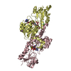
| Entry | Database: PDB / ID: 5dna | ||||||
|---|---|---|---|---|---|---|---|
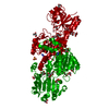
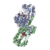
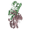
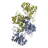
| Title | Crystal structure of Candida boidinii formate dehydrogenase | ||||||
 Components Components | FORMATE DEHYDROGENASE | ||||||
 Keywords Keywords | OXIDOREDUCTASE / isozyme / recombinant | ||||||
| Function / homology |  Function and homology information Function and homology informationformate catabolic process / formate dehydrogenase / formate dehydrogenase (NAD+) activity / oxidoreductase activity, acting on the CH-OH group of donors, NAD or NADP as acceptor / NAD binding / cytoplasmSimilarity search - Function | ||||||
| Biological species |  Candida boidinii (fungus) Candida boidinii (fungus) | ||||||
| Method | X-RAY DIFFRACTION / MOLECULAR REPLACEMENT / Resolution: 1.75 Å | ||||||
 Authors Authors | Guo, Q. / Gakhar, L. / Wichersham, K. / Francis, K. / Vardi-Kilshtain, A. / Major, D.T. / Cheatum, C.M. / Kohen, A. | ||||||
 Citation Citation | Journal: Biochemistry / Year: 2016 Title: Structural and Kinetic Studies of Formate Dehydrogenase from Candida boidinii. Authors: Guo, Q. / Gakhar, L. / Wickersham, K. / Francis, K. / Vardi-Kilshtain, A. / Major, D.T. / Cheatum, C.M. / Kohen, A. | ||||||
| History |
|
- Structure visualization
| Structure viewer | Molecule: MolmilJmol/JSmol |
|---|
- Downloads & links
-Download
| PDBx/mmCIF format | 5dna.cif.gz | 555.7 KB | Display | PDBx/mmCIF format |
|---|---|---|---|---|
| PDB format | pdb5dna.ent.gz | 458.5 KB | Display | PDB format |
| PDBx/mmJSON format | 5dna.json.gz | Tree view | PDBx/mmJSON format | |
| Others |  Other downloads Other downloads |
-Validation report
| Arichive directory | https://data.pdbj.org/pub/pdb/validation_reports/dn/5dnaftp://data.pdbj.org/pub/pdb/validation_reports/dn/5dna | HTTPS FTP |
|---|
-Related structure data
| Related structure data | 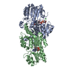 5dn9C 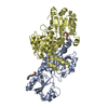 2j6iS C: citing same article ( S: Starting model for refinement |
|---|---|
| Similar structure data |


-Links
 PDBj
PDBj- Assembly
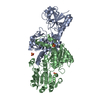
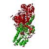
| Deposited unit | 
| ||||||||
|---|---|---|---|---|---|---|---|---|---|
| 1 | 
| ||||||||
| 2 | 
| ||||||||
| Unit cell |
|
-Components
| #1: Protein | Mass: 40364.969 Da / Num. of mol.: 4 Source method: isolated from a genetically manipulated source Source: (gene. exp.) Candida boidinii (fungus) / Gene: FDHProduction host:  Escherichia coli 'BL21-Gold(DE3)pLysS AG' (bacteria) Escherichia coli 'BL21-Gold(DE3)pLysS AG' (bacteria)References: UniProt: A0A0A1EQY0, formate dehydrogenase#2: Chemical | ChemComp-SO4 / Sulfate  Mass: 96.063 Da / Num. of mol.: 5 / Source method: obtained synthetically / Formula: SO4 Mass: 96.063 Da / Num. of mol.: 5 / Source method: obtained synthetically / Formula: SO4#3: Water | ChemComp-HOH / | Water Mass: 18.015 Da / Num. of mol.: 1777 / Source method: isolated from a natural source / Formula: H2O Mass: 18.015 Da / Num. of mol.: 1777 / Source method: isolated from a natural source / Formula: H2O |
|---|
-Experimental details
-Experiment
| Experiment | Method: X-RAY DIFFRACTION |
|---|
- Sample preparation
| Crystal | Density Matthews: 2.41 Å3/Da / Density % sol: 49 % |
|---|---|
| Crystal grow | Temperature: 291 K / Method: vapor diffusion, hanging drop / pH: 7.5 Details: 0.1 M sodium acetate trihydrate, 0.1 M HEPES, 25% PEG 4000, pH 7.5 |
-Data collection
| Diffraction | Mean temperature: 100 K |
|---|---|
| Diffraction source | Source: ROTATING ANODE / Type: RIGAKU RUH3R / Wavelength: 1.54 Å |
| Detector | Type: RIGAKU RAXIS IV++ / Detector: IMAGE PLATE / Date: Apr 3, 2012 |
| Radiation | Protocol: SINGLE WAVELENGTH / Monochromatic (M) / Laue (L): M / Scattering type: x-ray |
| Radiation wavelength | Wavelength: 1.54 Å / Relative weight: 1 |
| Reflection | Resolution: 1.75→19.9 Å / Num. obs: 141899 / % possible obs: 93.2 % / Redundancy: 3.8 % / Rmerge(I) obs: 0.061 / Net I/σ(I): 15.3 |
| Reflection shell | Resolution: 1.75→1.84 Å / Rmerge(I) obs: 0.411 / Mean I/σ(I) obs: 2.9 / % possible all: 90 |
- Processing
| Software |
| |||||||||||||||||||||||||||||||||||||||||||||||||||||||||||||||||||||||||||||||||||||||||||||||||||||||||||||||||||||||||||||||||||||||||||||||||||||||||||||||||||||||||||||||||||||||||||||||||||||||||||||||||||||||||
|---|---|---|---|---|---|---|---|---|---|---|---|---|---|---|---|---|---|---|---|---|---|---|---|---|---|---|---|---|---|---|---|---|---|---|---|---|---|---|---|---|---|---|---|---|---|---|---|---|---|---|---|---|---|---|---|---|---|---|---|---|---|---|---|---|---|---|---|---|---|---|---|---|---|---|---|---|---|---|---|---|---|---|---|---|---|---|---|---|---|---|---|---|---|---|---|---|---|---|---|---|---|---|---|---|---|---|---|---|---|---|---|---|---|---|---|---|---|---|---|---|---|---|---|---|---|---|---|---|---|---|---|---|---|---|---|---|---|---|---|---|---|---|---|---|---|---|---|---|---|---|---|---|---|---|---|---|---|---|---|---|---|---|---|---|---|---|---|---|---|---|---|---|---|---|---|---|---|---|---|---|---|---|---|---|---|---|---|---|---|---|---|---|---|---|---|---|---|---|---|---|---|---|---|---|---|---|---|---|---|---|---|---|---|---|---|---|---|---|
| Refinement | Method to determine structure: MOLECULAR REPLACEMENT Starting model: 2J6I Resolution: 1.75→19.895 Å / SU ML: 0.17 / Cross valid method: FREE R-VALUE / σ(F): 1.97 / Phase error: 18.15 / Stereochemistry target values: ML
| |||||||||||||||||||||||||||||||||||||||||||||||||||||||||||||||||||||||||||||||||||||||||||||||||||||||||||||||||||||||||||||||||||||||||||||||||||||||||||||||||||||||||||||||||||||||||||||||||||||||||||||||||||||||||
| Solvent computation | Shrinkage radii: 0.9 Å / VDW probe radii: 1.11 Å / Solvent model: FLAT BULK SOLVENT MODEL | |||||||||||||||||||||||||||||||||||||||||||||||||||||||||||||||||||||||||||||||||||||||||||||||||||||||||||||||||||||||||||||||||||||||||||||||||||||||||||||||||||||||||||||||||||||||||||||||||||||||||||||||||||||||||
| Refinement step | Cycle: LAST / Resolution: 1.75→19.895 Å
| |||||||||||||||||||||||||||||||||||||||||||||||||||||||||||||||||||||||||||||||||||||||||||||||||||||||||||||||||||||||||||||||||||||||||||||||||||||||||||||||||||||||||||||||||||||||||||||||||||||||||||||||||||||||||
| Refine LS restraints |
| |||||||||||||||||||||||||||||||||||||||||||||||||||||||||||||||||||||||||||||||||||||||||||||||||||||||||||||||||||||||||||||||||||||||||||||||||||||||||||||||||||||||||||||||||||||||||||||||||||||||||||||||||||||||||
| LS refinement shell |
|
