 Movie
Movie Controller

[English] 日本語
 Yorodumi
- PDB-5dch: Crystal structure of Pseudomonas aeruginosa DsbA E82I in complex ... -
+ Open data

- Basic information
| Entry | Database: PDB / ID: 5dch | |||||||||
|---|---|---|---|---|---|---|---|---|---|---|
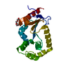
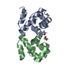
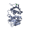
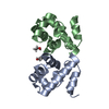
| Title | Crystal structure of Pseudomonas aeruginosa DsbA E82I in complex with MIPS-0000851 (3-[(2-METHYLBENZYL)SULFANYL]-4H-1,2,4-TRIAZOL-4-AMINE) | |||||||||
 Components Components | Thiol:disulfide interchange protein DsbA | |||||||||
 Keywords Keywords |  OXIDOREDUCTASE / thioredoxin fold OXIDOREDUCTASE / thioredoxin fold | |||||||||
| Function / homology |  Function and homology information Function and homology information | |||||||||
| Biological species |   Pseudomonas aeruginosa (bacteria) Pseudomonas aeruginosa (bacteria) | |||||||||
| Method | X-RAY DIFFRACTION / SYNCHROTRON / MOLECULAR REPLACEMENT / Resolution: 1.447 Å | |||||||||
 Authors Authors | McMahon, R.M. / Martin, J.L. | |||||||||
| Funding support |  Australia, 2items Australia, 2items
| |||||||||
 Citation Citation | Journal: PLoS ONE / Year: 2017 Title: Fragment library screening identifies hits that bind to the non-catalytic surface of Pseudomonas aeruginosa DsbA1. Authors: Mohanty, B. / Rimmer, K. / McMahon, R.M. / Headey, S.J. / Vazirani, M. / Shouldice, S.R. / Coincon, M. / Tay, S. / Morton, C.J. / Simpson, J.S. / Martin, J.L. / Scanlon, M.J. | |||||||||
| History |
|
- Structure visualization
| Structure viewer | Molecule: MolmilJmol/JSmol |
|---|
- Downloads & links
-Download
| PDBx/mmCIF format | 5dch.cif.gz | 129.4 KB | Display | PDBx/mmCIF format |
|---|---|---|---|---|
| PDB format | pdb5dch.ent.gz | 101.5 KB | Display | PDB format |
| PDBx/mmJSON format | 5dch.json.gz | Tree view | PDBx/mmJSON format | |
| Others |  Other downloads Other downloads |
-Validation report
| Arichive directory | https://data.pdbj.org/pub/pdb/validation_reports/dc/5dchftp://data.pdbj.org/pub/pdb/validation_reports/dc/5dch | HTTPS FTP |
|---|
-Related structure data
| Related structure data |  2mbtC  5tlqC  3h93S C: citing same article ( S: Starting model for refinement |
|---|---|
| Similar structure data |


-Links
 PDBj
PDBj

- Assembly
| Deposited unit | 
| ||||||||
|---|---|---|---|---|---|---|---|---|---|
| 1 |
| ||||||||
| Unit cell |
|
-Components
| #1: Protein | Mass: 21351.514 Da / Num. of mol.: 1 / Mutation: E82I Source method: isolated from a genetically manipulated source Source: (gene. exp.) Pseudomonas aeruginosa (bacteria) / Strain: ATCC 15692 / PAO1 / 1C / PRS 101 / LMG 12228 / Gene: dsbA, PA5489 / Plasmid: pET28a / Production host: Escherichia coli (E. coli) / Strain (production host): BL21(DE3) pLysS / References: UniProt: P0C2B2 | ||||
|---|---|---|---|---|---|
| #2: Chemical | ChemComp-1YO /   Mass: 220.294 Da / Num. of mol.: 1 / Source method: obtained synthetically / Formula: C10H12N4S Mass: 220.294 Da / Num. of mol.: 1 / Source method: obtained synthetically / Formula: C10H12N4S | ||||
| #3: Chemical | ChemComp-GOL / Glycerol  Mass: 92.094 Da / Num. of mol.: 4 / Source method: obtained synthetically / Formula: C3H8O3 Mass: 92.094 Da / Num. of mol.: 4 / Source method: obtained synthetically / Formula: C3H8O3#4: Chemical | ChemComp-MES / | MES (buffer)  Mass: 195.237 Da / Num. of mol.: 1 / Source method: obtained synthetically / Formula: C6H13NO4S / Comment: pH buffer*YM Mass: 195.237 Da / Num. of mol.: 1 / Source method: obtained synthetically / Formula: C6H13NO4S / Comment: pH buffer*YM#5: Water | ChemComp-HOH / | Water Mass: 18.015 Da / Num. of mol.: 141 / Source method: isolated from a natural source / Formula: H2O Mass: 18.015 Da / Num. of mol.: 141 / Source method: isolated from a natural source / Formula: H2O |
-Experimental details
-Experiment
| Experiment | Method: X-RAY DIFFRACTION / Number of used crystals: 1 |
|---|
- Sample preparation
| Crystal | Density Matthews: 2.23 Å3/Da / Density % sol: 44.73 % |
|---|---|
| Crystal grow | Temperature: 298 K / Method: vapor diffusion, hanging drop / pH: 6 / Details: 20-29 % PEG 1500, 0.1 M MES |
-Data collection
| Diffraction | Mean temperature: 100 K | |||||||||||||||||||||||||||
|---|---|---|---|---|---|---|---|---|---|---|---|---|---|---|---|---|---|---|---|---|---|---|---|---|---|---|---|---|
| Diffraction source | Source: SYNCHROTRON / Site: Australian Synchrotron / Beamline: MX2 / Wavelength: 0.9537 Å | |||||||||||||||||||||||||||
| Detector | Type: ADSC QUANTUM 315r / Detector: CCD / Date: Jul 26, 2013 | |||||||||||||||||||||||||||
| Radiation | Protocol: SINGLE WAVELENGTH / Monochromatic (M) / Laue (L): M / Scattering type: x-ray | |||||||||||||||||||||||||||
| Radiation wavelength | Wavelength: 0.9537 Å / Relative weight: 1 | |||||||||||||||||||||||||||
| Reflection | Resolution: 1.447→35.1 Å / Num. obs: 31349 / % possible obs: 96.4 % / Redundancy: 7.4 % / Biso Wilson estimate: 15.38 Å2 / CC1/2: 0.998 / Rmerge(I) obs: 0.082 / Rpim(I) all: 0.032 / Net I/σ(I): 14.1 / Num. measured all: 232922 / Scaling rejects: 12 | |||||||||||||||||||||||||||
| Reflection shell | Diffraction-ID: 1 / Rejects: 0
|
- Processing
| Software |
| ||||||||||||||||||||||||||||||||||||||||||||||||||||||||||||||||||||||||||||||||||||
|---|---|---|---|---|---|---|---|---|---|---|---|---|---|---|---|---|---|---|---|---|---|---|---|---|---|---|---|---|---|---|---|---|---|---|---|---|---|---|---|---|---|---|---|---|---|---|---|---|---|---|---|---|---|---|---|---|---|---|---|---|---|---|---|---|---|---|---|---|---|---|---|---|---|---|---|---|---|---|---|---|---|---|---|---|---|
| Refinement | Method to determine structure: MOLECULAR REPLACEMENT Starting model: 3H93 Resolution: 1.447→35.098 Å / FOM work R set: 0.9076 / SU ML: 0.1 / Cross valid method: FREE R-VALUE / σ(F): 1.34 / Phase error: 16.25 / Stereochemistry target values: ML
| ||||||||||||||||||||||||||||||||||||||||||||||||||||||||||||||||||||||||||||||||||||
| Solvent computation | Shrinkage radii: 0.9 Å / VDW probe radii: 1.11 Å / Solvent model: FLAT BULK SOLVENT MODEL | ||||||||||||||||||||||||||||||||||||||||||||||||||||||||||||||||||||||||||||||||||||
| Displacement parameters | Biso max: 66.58 Å2 / Biso mean: 19.19 Å2 / Biso min: 6.87 Å2 | ||||||||||||||||||||||||||||||||||||||||||||||||||||||||||||||||||||||||||||||||||||
| Refinement step | Cycle: final / Resolution: 1.447→35.098 Å
| ||||||||||||||||||||||||||||||||||||||||||||||||||||||||||||||||||||||||||||||||||||
| Refine LS restraints |
| ||||||||||||||||||||||||||||||||||||||||||||||||||||||||||||||||||||||||||||||||||||
| LS refinement shell | Refine-ID: X-RAY DIFFRACTION / Total num. of bins used: 11
|
