 Movie
Movie Controller


+ Open data

- Basic information
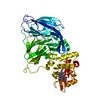
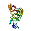
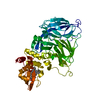
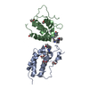
| Entry | Database: PDB / ID: 5c9v | |||||||||
|---|---|---|---|---|---|---|---|---|---|---|
| Title | Structure of human Parkin G319A | |||||||||
 Components Components | E3 ubiquitin-protein ligase parkin | |||||||||
 Keywords Keywords |  SIGNALING PROTEIN / Parkin / ubiquitin / E3 ligase / RBR / Parkinson's disease / mitophagy / cell signalling SIGNALING PROTEIN / Parkin / ubiquitin / E3 ligase / RBR / Parkinson's disease / mitophagy / cell signalling | |||||||||
| Function / homology |  Function and homology information Function and homology informationpositive regulation of retrograde transport, endosome to Golgi / regulation of lipid transport / positive regulation of neurotransmitter uptake / regulation protein catabolic process at presynapse / negative regulation of endoplasmic reticulum stress-induced neuron intrinsic apoptotic signaling pathway / negative regulation of primary amine oxidase activity / negative regulation of spontaneous neurotransmitter secretion / negative regulation of intralumenal vesicle formation / regulation of protein targeting to mitochondrion / negative regulation of glucokinase activity ...positive regulation of retrograde transport, endosome to Golgi / regulation of lipid transport / positive regulation of neurotransmitter uptake / regulation protein catabolic process at presynapse / negative regulation of endoplasmic reticulum stress-induced neuron intrinsic apoptotic signaling pathway / negative regulation of primary amine oxidase activity / negative regulation of spontaneous neurotransmitter secretion / negative regulation of intralumenal vesicle formation / regulation of protein targeting to mitochondrion / negative regulation of glucokinase activity / mitochondrion to lysosome vesicle-mediated transport / negative regulation of exosomal secretion / positive regulation of mitochondrial fusion / parkin-mediated stimulation of mitophagy in response to mitochondrial depolarization / protein K29-linked ubiquitination / Lewy body / protein K27-linked ubiquitination / Parkin-FBXW7-Cul1 ubiquitin ligase complex / free ubiquitin chain polymerization / negative regulation of actin filament bundle assembly / regulation of synaptic vesicle transport / negative regulation of mitochondrial fusion / RBR-type E3 ubiquitin transferase / positive regulation of mitophagy in response to mitochondrial depolarization / positive regulation of protein linear polyubiquitination / F-box domain binding / negative regulation by host of viral genome replication / positive regulation of mitophagy / dopaminergic synapse / cellular response to toxic substance / regulation of dopamine metabolic process / regulation of necroptotic process / regulation of cellular response to oxidative stress / negative regulation of intrinsic apoptotic signaling pathway by p53 class mediator / protein K6-linked ubiquitination / positive regulation of dendrite extension / norepinephrine metabolic process / positive regulation of proteasomal protein catabolic process / protein localization to mitochondrion / negative regulation of oxidative stress-induced neuron intrinsic apoptotic signaling pathway / positive regulation of protein localization to membrane / negative regulation of JNK cascade / protein K11-linked ubiquitination / positive regulation of tumor necrosis factor-mediated signaling pathway / cellular response to dopamine / autophagy of mitochondrion / mitochondrial fission / aggresome assembly / ubiquitin conjugating enzyme binding / regulation of canonical Wnt signaling pathway / ERAD pathway / aggresome / regulation of mitochondrion organization / regulation of reactive oxygen species metabolic process / dopamine uptake involved in synaptic transmission / regulation of synaptic vesicle endocytosis / positive regulation of mitochondrial fission / dopamine metabolic process / regulation of dopamine secretion / ubiquitin-specific protease binding / protein monoubiquitination / startle response / negative regulation of release of cytochrome c from mitochondria / protein K63-linked ubiquitination / cullin family protein binding / phospholipase binding / mitophagy / regulation of protein ubiquitination / regulation of glucose metabolic process / negative regulation of insulin secretion / protein K48-linked ubiquitination / negative regulation of reactive oxygen species metabolic process / protein autoubiquitination / positive regulation of DNA binding / cellular response to unfolded protein / cellular response to manganese ion / ubiquitin ligase complex / negative regulation of endoplasmic reticulum stress-induced intrinsic apoptotic signaling pathway / Hsp70 protein binding / heat shock protein binding / mitochondrion organization / PINK1-PRKN Mediated Mitophagy / response to endoplasmic reticulum stress / tubulin binding / adult locomotory behavior / Josephin domain DUBs / regulation of mitochondrial membrane potential / negative regulation of protein phosphorylation / ubiquitin binding / learning / synaptic transmission, glutamatergic / central nervous system development / regulation of autophagy / G protein-coupled receptor binding / PDZ domain binding / proteasomal protein catabolic process / macroautophagy / negative regulation of canonical Wnt signaling pathway / protein destabilization / regulation of protein stabilitySimilarity search - Function | |||||||||
| Biological species |  Homo sapiens (human) Homo sapiens (human) | |||||||||
| Method | X-RAY DIFFRACTION / SYNCHROTRON / MOLECULAR REPLACEMENT / Resolution: 2.35 Å | |||||||||
 Authors Authors | Wauer, T. / Komander, D. | |||||||||
| Funding support |  United Kingdom, 2items United Kingdom, 2items
| |||||||||
 Citation Citation | Journal: Nature / Year: 2015 Title: Mechanism of phospho-ubiquitin-induced PARKIN activation. Authors: Wauer, T. / Simicek, M. / Schubert, A. / Komander, D. #1: Journal: EMBO J. / Year: 2013Title: Structure of the human Parkin ligase domain in an autoinhibited state. Authors: Wauer, T. / Komander, D. | |||||||||
| History |
|
- Structure visualization
| Structure viewer | Molecule: MolmilJmol/JSmol |
|---|
- Downloads & links
-Download
| PDBx/mmCIF format | 5c9v.cif.gz | 138.6 KB | Display | PDBx/mmCIF format |
|---|---|---|---|---|
| PDB format | pdb5c9v.ent.gz | 107 KB | Display | PDB format |
| PDBx/mmJSON format | 5c9v.json.gz | Tree view | PDBx/mmJSON format | |
| Others |  Other downloads Other downloads |
-Validation report
| Arichive directory | https://data.pdbj.org/pub/pdb/validation_reports/c9/5c9vftp://data.pdbj.org/pub/pdb/validation_reports/c9/5c9v | HTTPS FTP |
|---|
-Related structure data
| Related structure data |  5cawC 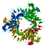 4bm9S S: Starting model for refinement C: citing same article ( |
|---|---|
| Similar structure data |


-Links
 PDBj
PDBj

- Assembly
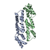
| Deposited unit | 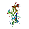
| |||||||||||||||
|---|---|---|---|---|---|---|---|---|---|---|---|---|---|---|---|---|
| 1 |
| |||||||||||||||
| Unit cell |
| |||||||||||||||
| Components on special symmetry positions |
|
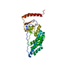
-Components
| #1: Protein | Mass: 36892.129 Da / Num. of mol.: 1 / Fragment: UNP residues 137-465 / Mutation: G319A Source method: isolated from a genetically manipulated source Details: engineered mutation at position G319A / Source: (gene. exp.) Homo sapiens (human) / Gene: PARK2, PRKN / Plasmid: pOPINKProduction host:  Escherichia coli 'BL21-Gold(DE3)pLysS AG' (bacteria) Escherichia coli 'BL21-Gold(DE3)pLysS AG' (bacteria)References: UniProt: O60260, Ligases; Forming carbon-nitrogen bonds; Acid-amino-acid ligases (peptide synthases) | ||||||
|---|---|---|---|---|---|---|---|
| #2: Chemical | ChemComp-ZN /   Mass: 65.409 Da / Num. of mol.: 8 / Source method: obtained synthetically / Formula: Zn Mass: 65.409 Da / Num. of mol.: 8 / Source method: obtained synthetically / Formula: Zn#3: Chemical | ChemComp-SO4 / Sulfate  Mass: 96.063 Da / Num. of mol.: 6 / Source method: obtained synthetically / Formula: SO4 Mass: 96.063 Da / Num. of mol.: 6 / Source method: obtained synthetically / Formula: SO4#4: Chemical | Glycerol  Mass: 92.094 Da / Num. of mol.: 3 / Source method: obtained synthetically / Formula: C3H8O3 Mass: 92.094 Da / Num. of mol.: 3 / Source method: obtained synthetically / Formula: C3H8O3#5: Water | ChemComp-HOH / | Water Mass: 18.015 Da / Num. of mol.: 81 / Source method: isolated from a natural source / Formula: H2O Mass: 18.015 Da / Num. of mol.: 81 / Source method: isolated from a natural source / Formula: H2O |
-Experimental details
-Experiment
| Experiment | Method: X-RAY DIFFRACTION |
|---|
- Sample preparation
| Crystal | Density Matthews: 3.63 Å3/Da / Density % sol: 66.1 % |
|---|---|
| Crystal grow | Temperature: 291 K / Method: vapor diffusion, sitting drop / pH: 5.6 Details: 1.8 M lithium sulphate, 0.01 M MgCl2, 0.05 M MES pH 5.6 PH range: 5.6 |
-Data collection
| Diffraction | Mean temperature: 100 K |
|---|---|
| Diffraction source | Source: SYNCHROTRON / Site: Diamond / Beamline: I04-1 / Wavelength: 0.9173 Å |
| Detector | Type: DECTRIS PILATUS 6M-F / Detector: PIXEL / Date: Apr 20, 2015 |
| Radiation | Protocol: SINGLE WAVELENGTH / Monochromatic (M) / Laue (L): M / Scattering type: x-ray |
| Radiation wavelength | Wavelength: 0.9173 Å / Relative weight: 1 |
| Reflection | Resolution: 2.35→86.68 Å / Num. obs: 22270 / % possible obs: 100 % / Observed criterion σ(I): 2 / Redundancy: 6.9 % / Biso Wilson estimate: 42.5 Å2 / Rmerge(I) obs: 0.089 / Net I/σ(I): 13.3 |
| Reflection shell | Resolution: 2.35→2.43 Å / Redundancy: 6.8 % / Rmerge(I) obs: 0.8 / Mean I/σ(I) obs: 2.1 / % possible all: 100 |
- Processing
| Software |
| |||||||||||||||||||||||||||||||||||||||||||||||||||||||||||||||
|---|---|---|---|---|---|---|---|---|---|---|---|---|---|---|---|---|---|---|---|---|---|---|---|---|---|---|---|---|---|---|---|---|---|---|---|---|---|---|---|---|---|---|---|---|---|---|---|---|---|---|---|---|---|---|---|---|---|---|---|---|---|---|---|---|
| Refinement | Method to determine structure: MOLECULAR REPLACEMENT Starting model: 4bm9 Resolution: 2.35→84.68 Å / SU ML: 0.22 / Cross valid method: FREE R-VALUE / σ(F): 1.33 / Phase error: 23.8 / Stereochemistry target values: ML
| |||||||||||||||||||||||||||||||||||||||||||||||||||||||||||||||
| Solvent computation | Shrinkage radii: 0.9 Å / VDW probe radii: 1.11 Å / Solvent model: FLAT BULK SOLVENT MODEL | |||||||||||||||||||||||||||||||||||||||||||||||||||||||||||||||
| Displacement parameters | Biso mean: 54.3 Å2 | |||||||||||||||||||||||||||||||||||||||||||||||||||||||||||||||
| Refinement step | Cycle: LAST / Resolution: 2.35→84.68 Å
| |||||||||||||||||||||||||||||||||||||||||||||||||||||||||||||||
| Refine LS restraints |
| |||||||||||||||||||||||||||||||||||||||||||||||||||||||||||||||
| LS refinement shell |
| |||||||||||||||||||||||||||||||||||||||||||||||||||||||||||||||
| Refinement TLS params. | Method: refined / Origin x: 7.8845 Å / Origin y: 31.4194 Å / Origin z: 36.9402 Å
| |||||||||||||||||||||||||||||||||||||||||||||||||||||||||||||||
| Refinement TLS group | Selection details: chain A |
