 Movie
Movie Controller

[English] 日本語
 Yorodumi
- PDB-5bpk: Varying binding modes of inhibitors and structural differences in... -
+ Open data

- Basic information
| Entry | Database: PDB / ID: 5bpk | ||||||
|---|---|---|---|---|---|---|---|
| Title | Varying binding modes of inhibitors and structural differences in the binding pockets of different gamma-glutamyltranspeptidases | ||||||
 Components Components | (Gamma-glutamyltranspeptidase (Ggt)) x 2 | ||||||
 Keywords Keywords |  HYDROLASE / gamma-GLUTAMYLTRANSPEPTIDASE / NTN-HYDROLASE / Acivicin / PROTEROS BIOSTRUCTURES GMBH HYDROLASE / gamma-GLUTAMYLTRANSPEPTIDASE / NTN-HYDROLASE / Acivicin / PROTEROS BIOSTRUCTURES GMBH | ||||||
| Function / homology |  Function and homology informationgamma-glutamyltransferase / glutathione gamma-glutamate hydrolase / glutathione hydrolase activity / leukotriene C4 gamma-glutamyl transferase activity / glutathione catabolic process / negative regulation of cell cycle G1/S phase transition / glutathione biosynthetic process / negative regulation of T cell proliferation / positive regulation of interleukin-8 production Function and homology informationgamma-glutamyltransferase / glutathione gamma-glutamate hydrolase / glutathione hydrolase activity / leukotriene C4 gamma-glutamyl transferase activity / glutathione catabolic process / negative regulation of cell cycle G1/S phase transition / glutathione biosynthetic process / negative regulation of T cell proliferation / positive regulation of interleukin-8 productionSimilarity search - Function | ||||||
| Biological species |  Helicobacter pylori 26695 (bacteria) Helicobacter pylori 26695 (bacteria) Helicobacter pylori (bacteria) Helicobacter pylori (bacteria) | ||||||
| Method | X-RAY DIFFRACTION / SYNCHROTRON / MOLECULAR REPLACEMENT / molecular replacement / Resolution: 1.49 Å | ||||||
 Authors Authors | Bolz, C. / Bach, N.C. / Meyer, H. / Mueller, G. / Dawidowski, M. / Popowicz, G. / Sieber, S.A. / Skerra, A. / Gerhard, M. | ||||||
 Citation Citation | Journal: To Be Published Title: Varying binding modes of inhibitors and structural differences in the binding pockets of different gamma-glutamyltranspeptidases Authors: Bolz, C. / Bach, N.C. / Meyer, H. / Mueller, G. / Dawidowski, M. / Popowicz, G. / Sieber, S.A. / Skerra, A. / Gerhard, M. | ||||||
| History |
|
- Structure visualization
| Structure viewer | Molecule: MolmilJmol/JSmol |
|---|
- Downloads & links
-Download
| PDBx/mmCIF format | 5bpk.cif.gz | 463.6 KB | Display | PDBx/mmCIF format |
|---|---|---|---|---|
| PDB format | pdb5bpk.ent.gz | 376.8 KB | Display | PDB format |
| PDBx/mmJSON format | 5bpk.json.gz | Tree view | PDBx/mmJSON format | |
| Others |  Other downloads Other downloads |
-Validation report
| Arichive directory | https://data.pdbj.org/pub/pdb/validation_reports/bp/5bpkftp://data.pdbj.org/pub/pdb/validation_reports/bp/5bpk | HTTPS FTP |
|---|
-Related structure data
| Related structure data |  2nqoS S: Starting model for refinement |
|---|---|
| Similar structure data |


-Links
 PDBj
PDBj


- Assembly
| Deposited unit | 
| ||||||||
|---|---|---|---|---|---|---|---|---|---|
| 1 | 
| ||||||||
| 2 | 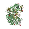
| ||||||||
| Unit cell |
|
-Components
| #1: Protein | Mass: 40832.039 Da / Num. of mol.: 2 / Fragment: CATALYTIC DOMAIN, residues 1-379 Source method: isolated from a genetically manipulated source Source: (gene. exp.) Helicobacter pylori 26695 (bacteria) / Gene: HP_1118 / Production host: Escherichia coli (E. coli) / References: UniProt: O25743#2: Protein | Mass: 24557.641 Da / Num. of mol.: 2 / Fragment: CATALYTIC DOMAIN, residues 380-567 Source method: isolated from a genetically manipulated source Source: (gene. exp.) Helicobacter pylori 26695 (bacteria) / Strain: / 26695 / Gene: HP_1118 / Production host: Escherichia coli (E. coli) / References: UniProt: O25743#3: Chemical | ChemComp-EDO / Ethylene glycol  Mass: 62.068 Da / Num. of mol.: 43 / Source method: obtained synthetically / Formula: C2H6O2 Mass: 62.068 Da / Num. of mol.: 43 / Source method: obtained synthetically / Formula: C2H6O2#4: Chemical |   Type: L-peptide linking / Mass: 144.129 Da / Num. of mol.: 2 Type: L-peptide linking / Mass: 144.129 Da / Num. of mol.: 2Source method: isolated from a genetically manipulated source Formula: C5H8N2O3 / Source: (gene. exp.) Helicobacter pylori (bacteria) / Production host: Escherichia coli (E. coli)#5: Water | ChemComp-HOH / | Water Mass: 18.015 Da / Num. of mol.: 958 / Source method: isolated from a natural source / Formula: H2O Mass: 18.015 Da / Num. of mol.: 958 / Source method: isolated from a natural source / Formula: H2O |
|---|
-Experimental details
-Experiment
| Experiment | Method: X-RAY DIFFRACTION / Number of used crystals: 1 |
|---|
- Sample preparation
| Crystal | Density Matthews: 2.82 Å3/Da / Density % sol: 56.4 % |
|---|---|
| Crystal grow | Temperature: 293 K / Method: vapor diffusion / Details: PEG3350, 0.1M TRIS |
-Data collection
| Diffraction | Mean temperature: 100 K |
|---|---|
| Diffraction source | Source: SYNCHROTRON / Site: SLS  / Beamline: X10SA / Wavelength: 1 Å / Beamline: X10SA / Wavelength: 1 Å |
| Detector | Type: MARRESEARCH / Detector: CCD / Date: Apr 26, 2010 |
| Radiation | Protocol: SINGLE WAVELENGTH / Monochromatic (M) / Laue (L): M / Scattering type: x-ray |
| Radiation wavelength | Wavelength: 1 Å / Relative weight: 1 |
| Reflection | Resolution: 1.49→91.84 Å / Num. obs: 176874 / % possible obs: 99.2 % / Observed criterion σ(I): 0 / Redundancy: 3.7 % / Rmerge(I) obs: 0.07 / Net I/σ(I): 16.6 |
| Reflection shell | Resolution: 1.49→1.55 Å / Redundancy: 3.7 % / Rmerge(I) obs: 0.51 / Rejects: 0 / % possible all: 98.5 |
-Phasing
| Phasing | Method: molecular replacement |
|---|
- Processing
| Software |
| ||||||||||||||||||||||||||||||||||||||||||||||||||||||||||||||||||||||||||||||||||||||||||||||||||||
|---|---|---|---|---|---|---|---|---|---|---|---|---|---|---|---|---|---|---|---|---|---|---|---|---|---|---|---|---|---|---|---|---|---|---|---|---|---|---|---|---|---|---|---|---|---|---|---|---|---|---|---|---|---|---|---|---|---|---|---|---|---|---|---|---|---|---|---|---|---|---|---|---|---|---|---|---|---|---|---|---|---|---|---|---|---|---|---|---|---|---|---|---|---|---|---|---|---|---|---|---|---|
| Refinement | Method to determine structure: MOLECULAR REPLACEMENT Starting model: 2NQO Resolution: 1.49→91.8 Å / Cor.coef. Fo:Fc: 0.97 / Cor.coef. Fo:Fc free: 0.954 / SU B: 2.592 / SU ML: 0.043 / Cross valid method: THROUGHOUT / σ(F): 0 / ESU R: 0.072 / ESU R Free: 0.065 / Stereochemistry target values: MAXIMUM LIKELIHOOD Details: HYDROGENS HAVE BEEN ADDED IN THE RIDING POSITIONS U VALUES : REFINED INDIVIDUALLY
| ||||||||||||||||||||||||||||||||||||||||||||||||||||||||||||||||||||||||||||||||||||||||||||||||||||
| Solvent computation | Ion probe radii: 0.8 Å / Shrinkage radii: 0.8 Å / VDW probe radii: 1.4 Å / Solvent model: BABINET MODEL WITH MASK | ||||||||||||||||||||||||||||||||||||||||||||||||||||||||||||||||||||||||||||||||||||||||||||||||||||
| Displacement parameters | Biso max: 57.14 Å2 / Biso mean: 13.238 Å2 / Biso min: 4.29 Å2
| ||||||||||||||||||||||||||||||||||||||||||||||||||||||||||||||||||||||||||||||||||||||||||||||||||||
| Refinement step | Cycle: final / Resolution: 1.49→91.8 Å
| ||||||||||||||||||||||||||||||||||||||||||||||||||||||||||||||||||||||||||||||||||||||||||||||||||||
| Refine LS restraints |
| ||||||||||||||||||||||||||||||||||||||||||||||||||||||||||||||||||||||||||||||||||||||||||||||||||||
| LS refinement shell | Resolution: 1.49→1.529 Å / Total num. of bins used: 20
|
