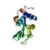
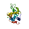
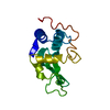
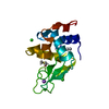
 Movie
Movie Controller


+ Open data

- Basic information
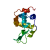
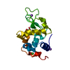
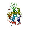
| Entry | Database: PDB / ID: 5b1g | ||||||
|---|---|---|---|---|---|---|---|
| Title | Crystal structure of hen egg-white lysozyme | ||||||
 Components Components | Lysozyme C | ||||||
 Keywords Keywords |  HYDROLASE / serial femtosecond crystallography HYDROLASE / serial femtosecond crystallography | ||||||
| Function / homology |  Function and homology informationAntimicrobial peptides / Neutrophil degranulation / beta-N-acetylglucosaminidase activity / cell wall macromolecule catabolic process / lysozyme / lysozyme activity / killing of cells of another organism / defense response to Gram-negative bacterium / defense response to Gram-positive bacterium / defense response to bacterium ...Antimicrobial peptides / Neutrophil degranulation / beta-N-acetylglucosaminidase activity / cell wall macromolecule catabolic process / lysozyme / lysozyme activity / killing of cells of another organism / defense response to Gram-negative bacterium / defense response to Gram-positive bacterium / defense response to bacterium / endoplasmic reticulum / extracellular space / identical protein binding / cytoplasm Function and homology informationAntimicrobial peptides / Neutrophil degranulation / beta-N-acetylglucosaminidase activity / cell wall macromolecule catabolic process / lysozyme / lysozyme activity / killing of cells of another organism / defense response to Gram-negative bacterium / defense response to Gram-positive bacterium / defense response to bacterium ...Antimicrobial peptides / Neutrophil degranulation / beta-N-acetylglucosaminidase activity / cell wall macromolecule catabolic process / lysozyme / lysozyme activity / killing of cells of another organism / defense response to Gram-negative bacterium / defense response to Gram-positive bacterium / defense response to bacterium / endoplasmic reticulum / extracellular space / identical protein binding / cytoplasmSimilarity search - Function | ||||||
| Biological species |  Gallus gallus (chicken) Gallus gallus (chicken) | ||||||
| Method | X-RAY DIFFRACTION / FREE ELECTRON LASER / MOLECULAR REPLACEMENT / Resolution: 2.3 Å | ||||||
 Authors Authors | Sugahara, M. / Suzuki, M. / Nango, E. | ||||||
 Citation Citation | Journal: To Be Published Title: Crystal structure of hen egg-white lysozyme Authors: Sugahara, M. / Suzuki, M. / Nango, E. | ||||||
| History |
|
- Structure visualization
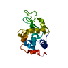
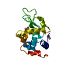
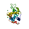
| Structure viewer | Molecule: MolmilJmol/JSmol |
|---|
- Downloads & links
-Download
| PDBx/mmCIF format | 5b1g.cif.gz | 39.6 KB | Display | PDBx/mmCIF format |
|---|---|---|---|---|
| PDB format | pdb5b1g.ent.gz | 25.3 KB | Display | PDB format |
| PDBx/mmJSON format | 5b1g.json.gz | Tree view | PDBx/mmJSON format | |
| Others |  Other downloads Other downloads |
-Validation report
| Arichive directory | https://data.pdbj.org/pub/pdb/validation_reports/b1/5b1gftp://data.pdbj.org/pub/pdb/validation_reports/b1/5b1g | HTTPS FTP |
|---|
-Related structure data
| Related structure data |  3wunC  4ym8C  5b1fC 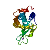 3wulS C: citing same article ( S: Starting model for refinement |
|---|---|
| Similar structure data | |
| Experimental dataset #1 | Data reference: 10.11577/1249397 / Data set type: diffraction image data |


-Links
 PDBj
PDBj


- Assembly
| Deposited unit | 
| ||||||||
|---|---|---|---|---|---|---|---|---|---|
| 1 |
| ||||||||
| Unit cell |
|
-Components
| #1: Protein | Mass: 14331.160 Da / Num. of mol.: 1 / Source method: isolated from a natural source / Source: (natural) Gallus gallus (chicken) / References: UniProt: P00698, lysozyme |
|---|---|
| #2: Chemical | ChemComp-NA /   Mass: 22.990 Da / Num. of mol.: 1 / Source method: obtained synthetically / Formula: Na Mass: 22.990 Da / Num. of mol.: 1 / Source method: obtained synthetically / Formula: Na |
| #3: Chemical | ChemComp-CL / Chloride  Mass: 35.453 Da / Num. of mol.: 1 / Source method: obtained synthetically / Formula: Cl Mass: 35.453 Da / Num. of mol.: 1 / Source method: obtained synthetically / Formula: Cl |
| #4: Water | ChemComp-HOH / Water Mass: 18.015 Da / Num. of mol.: 30 / Source method: isolated from a natural source / Formula: H2O Mass: 18.015 Da / Num. of mol.: 30 / Source method: isolated from a natural source / Formula: H2O |
-Experimental details
-Experiment
| Experiment | Method: X-RAY DIFFRACTION |
|---|
- Sample preparation
| Crystal | Density Matthews: 2.18 Å3/Da / Density % sol: 43.49 % |
|---|---|
| Crystal grow | Temperature: 293 K / Method: batch mode / Details: NaCl |
-Data collection
| Diffraction | Mean temperature: 300 K |
|---|---|
| Diffraction source | Source: FREE ELECTRON LASER / Site: SACLA  / Beamline: BL3 / Wavelength: 1.77 Å / Beamline: BL3 / Wavelength: 1.77 Å |
| Detector | Type: MPCCD / Detector: CCD / Date: Nov 25, 2014 |
| Radiation | Protocol: SINGLE WAVELENGTH / Monochromatic (M) / Laue (L): M / Scattering type: x-ray |
| Radiation wavelength | Wavelength: 1.77 Å / Relative weight: 1 |
| Reflection | Resolution: 2.3→30 Å / Num. obs: 5983 / % possible obs: 100 % / Redundancy: 502 % / Net I/σ(I): 9.8 |
| Reflection shell | Resolution: 2.3→2.34 Å / Mean I/σ(I) obs: 7.6 / % possible all: 100 |
- Processing
| Software |
| ||||||||||||||||||||||||
|---|---|---|---|---|---|---|---|---|---|---|---|---|---|---|---|---|---|---|---|---|---|---|---|---|---|
| Refinement | Method to determine structure: MOLECULAR REPLACEMENT Starting model: 3WUL Resolution: 2.3→28.39 Å / SU ML: 0.21 / Cross valid method: FREE R-VALUE / σ(F): 1.39 / Phase error: 20.39 / Stereochemistry target values: ML
| ||||||||||||||||||||||||
| Solvent computation | Shrinkage radii: 0.9 Å / VDW probe radii: 1.11 Å / Solvent model: FLAT BULK SOLVENT MODEL | ||||||||||||||||||||||||
| Refinement step | Cycle: LAST / Resolution: 2.3→28.39 Å
| ||||||||||||||||||||||||
| Refine LS restraints |
| ||||||||||||||||||||||||
| LS refinement shell |
|
