 Movie
Movie Controller


+ Open data

- Basic information
| Entry | Database: PDB / ID: 5ayl | ||||||
|---|---|---|---|---|---|---|---|
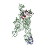
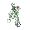
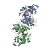
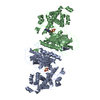
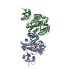
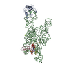
| Title | Crystal structure of ERdj5 form II | ||||||
 Components Components | DnaJ homolog subfamily C member 10 | ||||||
 Keywords Keywords |  OXIDOREDUCTASE / PDI FAMILY / THIOREDOXIN / ENDOPLASMIC RETICULUM OXIDOREDUCTASE / PDI FAMILY / THIOREDOXIN / ENDOPLASMIC RETICULUM | ||||||
| Function / homology |  Function and homology information Function and homology informationoxidoreductase activity, acting on a sulfur group of donors, disulfide as acceptor / Oxidoreductases; Acting on a sulfur group of donors; With a disulfide as acceptor / endoplasmic reticulum chaperone complex / protein folding in endoplasmic reticulum / disulfide oxidoreductase activity / misfolded protein binding / positive regulation of ATP-dependent activity / IRE1-mediated unfolded protein response / ATPase activator activity / protein-disulfide reductase activity ...oxidoreductase activity, acting on a sulfur group of donors, disulfide as acceptor / Oxidoreductases; Acting on a sulfur group of donors; With a disulfide as acceptor / endoplasmic reticulum chaperone complex / protein folding in endoplasmic reticulum / disulfide oxidoreductase activity / misfolded protein binding / positive regulation of ATP-dependent activity / IRE1-mediated unfolded protein response / ATPase activator activity / protein-disulfide reductase activity / intrinsic apoptotic signaling pathway in response to endoplasmic reticulum stress / : / Hsp70 protein binding / response to endoplasmic reticulum stress / negative regulation of protein phosphorylation / ATPase binding / protein-folding chaperone binding / endoplasmic reticulum lumen / endoplasmic reticulumSimilarity search - Function | ||||||
| Biological species |  Mus musculus (house mouse) Mus musculus (house mouse) | ||||||
| Method | X-RAY DIFFRACTION / SYNCHROTRON / MOLECULAR REPLACEMENT / Resolution: 2.4 Å | ||||||
 Authors Authors | Watanabe, S. / Maegawa, K. / Inaba, K. | ||||||
 Citation Citation | Journal: To Be Published Title: Highly dynamic nature of ERdj5 is essential for enhancement of the ER associated degradation Authors: Maegawa, K. / Watanabe, S. / Okumura, M. / Noi, K. / Inoue, M. / Ushioda, R. / Ogura, T. / Nagata, K. / Inaba, K. | ||||||
| History |
|
- Structure visualization
| Structure viewer | Molecule: MolmilJmol/JSmol |
|---|
- Downloads & links
-Download
| PDBx/mmCIF format | 5ayl.cif.gz | 312.1 KB | Display | PDBx/mmCIF format |
|---|---|---|---|---|
| PDB format | pdb5ayl.ent.gz | 249.7 KB | Display | PDB format |
| PDBx/mmJSON format | 5ayl.json.gz | Tree view | PDBx/mmJSON format | |
| Others |  Other downloads Other downloads |
-Validation report
| Arichive directory | https://data.pdbj.org/pub/pdb/validation_reports/ay/5aylftp://data.pdbj.org/pub/pdb/validation_reports/ay/5ayl | HTTPS FTP |
|---|
-Related structure data
| Related structure data | 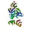 5aykC 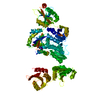 3apoS C: citing same article ( S: Starting model for refinement |
|---|---|
| Similar structure data |


-Links
 PDBj
PDBj


- Assembly
| Deposited unit | 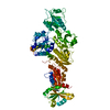
| ||||||||
|---|---|---|---|---|---|---|---|---|---|
| 1 |
| ||||||||
| Unit cell |
|
-Components
| #1: Protein | Mass: 89317.570 Da / Num. of mol.: 1 / Fragment: UNP residues 32-793 Mutation: C148S, C151S, G389V, C409S, C470S, C473S, C578S, C581S, C690S, C693S Source method: isolated from a genetically manipulated source Source: (gene. exp.) Mus musculus (house mouse) / Gene: Dnajc10, Erdj5, Jpdi / Plasmid: pET15b / Production host:  Escherichia coli (E. coli) / Strain (production host): Origami Escherichia coli (E. coli) / Strain (production host): OrigamiReferences: UniProt: Q9DC23, Oxidoreductases; Acting on a sulfur group of donors; With a disulfide as acceptor |
|---|---|
| #2: Chemical | ChemComp-1PS /   Mass: 201.243 Da / Num. of mol.: 1 / Source method: obtained synthetically / Formula: C8H11NO3S Mass: 201.243 Da / Num. of mol.: 1 / Source method: obtained synthetically / Formula: C8H11NO3S |
| #3: Water | ChemComp-HOH / Water Mass: 18.015 Da / Num. of mol.: 200 / Source method: isolated from a natural source / Formula: H2O Mass: 18.015 Da / Num. of mol.: 200 / Source method: isolated from a natural source / Formula: H2O |
-Experimental details
-Experiment
| Experiment | Method: X-RAY DIFFRACTION |
|---|
- Sample preparation
| Crystal | Density Matthews: 2.51 Å3/Da / Density % sol: 51.04 % Description: THE ENTRY CONTAINS FRIEDEL PAIRS IN F_PLUS/MINUS COLUMNS. |
|---|---|
| Crystal grow | Temperature: 293 K / Method: vapor diffusion, sitting drop / pH: 7.5 / Details: PEG8000, cystin / PH range: 7.5 or 8.2 |
-Data collection
| Diffraction | Mean temperature: 100 K |
|---|---|
| Diffraction source | Source: SYNCHROTRON / Site: Photon Factory  / Beamline: BL-1A / Wavelength: 1.1 Å / Beamline: BL-1A / Wavelength: 1.1 Å |
| Detector | Type: DECTRIS PILATUS 2M-F / Detector: PIXEL / Date: Jan 22, 2014 |
| Radiation | Protocol: SINGLE WAVELENGTH / Monochromatic (M) / Laue (L): M / Scattering type: x-ray |
| Radiation wavelength | Wavelength: 1.1 Å / Relative weight: 1 |
| Reflection | Resolution: 2.4→50 Å / Num. obs: 68376 / % possible obs: 98 % / Redundancy: 3.2 % / Rsym value: 0.126 / Net I/σ(I): 9.1 |
| Reflection shell | Resolution: 2.4→2.46 Å / Rmerge(I) obs: 0.688 / Mean I/σ(I) obs: 1.2 / % possible all: 82.1 |
- Processing
| Software |
| ||||||||||||||||||||||||||||||||||||||||||||||||||||||||||||||||||||||||||||||||||||||||||||||||||||||||||||||||||||||||||||||||||||||||||||||||||||||||||||||||||||||||||||||||||||||||||||||||||||||||
|---|---|---|---|---|---|---|---|---|---|---|---|---|---|---|---|---|---|---|---|---|---|---|---|---|---|---|---|---|---|---|---|---|---|---|---|---|---|---|---|---|---|---|---|---|---|---|---|---|---|---|---|---|---|---|---|---|---|---|---|---|---|---|---|---|---|---|---|---|---|---|---|---|---|---|---|---|---|---|---|---|---|---|---|---|---|---|---|---|---|---|---|---|---|---|---|---|---|---|---|---|---|---|---|---|---|---|---|---|---|---|---|---|---|---|---|---|---|---|---|---|---|---|---|---|---|---|---|---|---|---|---|---|---|---|---|---|---|---|---|---|---|---|---|---|---|---|---|---|---|---|---|---|---|---|---|---|---|---|---|---|---|---|---|---|---|---|---|---|---|---|---|---|---|---|---|---|---|---|---|---|---|---|---|---|---|---|---|---|---|---|---|---|---|---|---|---|---|---|---|---|---|
| Refinement | Method to determine structure: MOLECULAR REPLACEMENT Starting model: 3APO Resolution: 2.4→39.085 Å / SU ML: 0.31 / Cross valid method: FREE R-VALUE / σ(F): 1.35 / Phase error: 25.34 / Stereochemistry target values: ML Details: SF FILE CONTAINS FRIEDEL PAIRS UNDER I/F_MINUS AND I/F_PLUS COLUMNS.
| ||||||||||||||||||||||||||||||||||||||||||||||||||||||||||||||||||||||||||||||||||||||||||||||||||||||||||||||||||||||||||||||||||||||||||||||||||||||||||||||||||||||||||||||||||||||||||||||||||||||||
| Solvent computation | Shrinkage radii: 0.9 Å / VDW probe radii: 1.11 Å / Solvent model: FLAT BULK SOLVENT MODEL | ||||||||||||||||||||||||||||||||||||||||||||||||||||||||||||||||||||||||||||||||||||||||||||||||||||||||||||||||||||||||||||||||||||||||||||||||||||||||||||||||||||||||||||||||||||||||||||||||||||||||
| Refinement step | Cycle: LAST / Resolution: 2.4→39.085 Å
| ||||||||||||||||||||||||||||||||||||||||||||||||||||||||||||||||||||||||||||||||||||||||||||||||||||||||||||||||||||||||||||||||||||||||||||||||||||||||||||||||||||||||||||||||||||||||||||||||||||||||
| Refine LS restraints |
| ||||||||||||||||||||||||||||||||||||||||||||||||||||||||||||||||||||||||||||||||||||||||||||||||||||||||||||||||||||||||||||||||||||||||||||||||||||||||||||||||||||||||||||||||||||||||||||||||||||||||
| LS refinement shell |
| ||||||||||||||||||||||||||||||||||||||||||||||||||||||||||||||||||||||||||||||||||||||||||||||||||||||||||||||||||||||||||||||||||||||||||||||||||||||||||||||||||||||||||||||||||||||||||||||||||||||||
| Refinement TLS params. | Method: refined / Refine-ID: X-RAY DIFFRACTION
| ||||||||||||||||||||||||||||||||||||||||||||||||||||||||||||||||||||||||||||||||||||||||||||||||||||||||||||||||||||||||||||||||||||||||||||||||||||||||||||||||||||||||||||||||||||||||||||||||||||||||
| Refinement TLS group |
|
