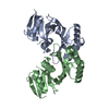
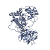
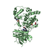
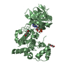
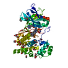
[English] 日本語
Yorodumi
- PDB-5awm: The Crystal Structure of JNK from Drosophila melanogaster Reveals... -

+
Open data


ID or keywords:

Loading...

-
Basic information

Entry
Database: PDB / ID: 5awm
TitleThe Crystal Structure of JNK from Drosophila melanogaster Reveals an Evolutionarily Conserved Topology with that of Mammalian JNK Proteins.
ComponentsStress-activated protein kinase JNK
KeywordsTRANSFERASE / c-Jun N-terminal kinase / MAP kinase / Drosophila JNK pathway
Function / homology
Function and homology information


collateral sprouting of injured axon / Formation of the cytosolic BSK 'scaffolding complex' / Formation of the nuclear AP-1 transcription factor 'scaffolding complex' / Transcriptional activtion by AP-1 transcription factor / Imd pathway / Activation of the AP-1 family of transcription factors / Interleukin-38 signaling / NRAGE signals death through JNK / chorion micropyle formation / embryonic anterior midgut (ectodermal) morphogenesis ...collateral sprouting of injured axon / Formation of the cytosolic BSK 'scaffolding complex' / Formation of the nuclear AP-1 transcription factor 'scaffolding complex' / Transcriptional activtion by AP-1 transcription factor / Imd pathway / Activation of the AP-1 family of transcription factors / Interleukin-38 signaling / NRAGE signals death through JNK / chorion micropyle formation / embryonic anterior midgut (ectodermal) morphogenesis / dorsal closure, spreading of leading edge cells / peptidoglycan recognition protein signaling pathway / imaginal disc fusion, thorax closure / imaginal disc-derived male genitalia morphogenesis / determination of digestive tract left/right asymmetry / FCERI mediated MAPK activation / Oxidative Stress Induced Senescence / JNK (c-Jun kinases) phosphorylation and activation mediated by activated human TAK1 / melanization defense response / positive regulation of neuron remodeling / dorsal closure / mushroom body development / follicle cell of egg chamber development / JUN phosphorylation / dorsal appendage formation / positive regulation of border follicle cell migration / JUN kinase activity / engulfment of apoptotic cell / cellular response to arsenic-containing substance / axon extension / wound healing, spreading of epidermal cells / neuron development / long-term memory / MAP kinase activity / mitogen-activated protein kinase / vascular endothelial growth factor receptor signaling pathway / positive regulation of autophagy / JNK cascade / cellular response to cadmium ion / tumor necrosis factor-mediated signaling pathway / neuron projection morphogenesis / axon guidance / wound healing / cellular response to reactive oxygen species / cellular response to oxidative stress / antibacterial humoral response / response to heat / regulation of gene expression / response to oxidative stress / protein kinase activity / intracellular signal transduction / axon / protein serine/threonine kinase activity / dendrite / positive regulation of gene expression / nucleoplasm / ATP binding / nucleus / cytosol / cytoplasm
Similarity search - Function
Mitogen-activated protein (MAP) kinase, JNK / Mitogen-activated protein (MAP) kinase, conserved site / MAP kinase signature. / Transferase(Phosphotransferase) domain 1 / Transferase(Phosphotransferase); domain 1 / Phosphorylase Kinase; domain 1 / Phosphorylase Kinase; domain 1 / Serine/threonine-protein kinase, active site / Serine/Threonine protein kinases active-site signature. / Protein kinase domain ...Mitogen-activated protein (MAP) kinase, JNK / Mitogen-activated protein (MAP) kinase, conserved site / MAP kinase signature. / Transferase(Phosphotransferase) domain 1 / Transferase(Phosphotransferase); domain 1 / Phosphorylase Kinase; domain 1 / Phosphorylase Kinase; domain 1 / Serine/threonine-protein kinase, active site / Serine/Threonine protein kinases active-site signature. / Protein kinase domain / Serine/Threonine protein kinases, catalytic domain / Protein kinase domain profile. / Protein kinase domain / Protein kinase-like domain superfamily / 2-Layer Sandwich / Orthogonal Bundle / Mainly Alpha / Alpha Beta
Similarity search - Domain/homology
PHOSPHOAMINOPHOSPHONIC ACID-ADENYLATE ESTER / Stress-activated protein kinase JNK
Similarity search - Component
Biological speciesDrosophila melanogaster (fruit fly)
MethodX-RAY DIFFRACTION / SYNCHROTRON / MOLECULAR REPLACEMENT / Resolution: 1.79 Å
AuthorsBoonserm, P.
Funding support Thailand, 1items
OrganizationGrant numberCountry
Mahidol University Thailand
CitationJournal: Bmc Struct.Biol. / Year: 2015
Title: The crystal structure of JNK from Drosophila melanogaster reveals an evolutionarily conserved topology with that of mammalian JNK proteins.
Authors: Chimnaronk, S. / Sitthiroongruang, J. / Srisucharitpanit, K. / Srisaisup, M. / Ketterman, A.J. / Boonserm, P.
History
DepositionJul 6, 2015Deposition site: PDBJ / Processing site: PDBJ
SupersessionAug 5, 2015ID: 4M3A
Revision 1.0Aug 5, 2015Provider: repository / Type: Initial release
Revision 1.1Nov 11, 2015Group: Database references
Revision 1.2Feb 26, 2020Group: Data collection / Derived calculations / Category: diffrn_source / pdbx_struct_oper_list
Item: _diffrn_source.pdbx_synchrotron_site / _pdbx_struct_oper_list.symmetry_operation
Revision 1.3Nov 8, 2023Group: Data collection / Database references ...Data collection / Database references / Derived calculations / Refinement description
Category: chem_comp_atom / chem_comp_bond ...chem_comp_atom / chem_comp_bond / database_2 / pdbx_initial_refinement_model / struct_conn
Item: _database_2.pdbx_DOI / _database_2.pdbx_database_accession ..._database_2.pdbx_DOI / _database_2.pdbx_database_accession / _struct_conn.pdbx_dist_value / _struct_conn.ptnr1_label_atom_id / _struct_conn.ptnr2_auth_seq_id / _struct_conn.ptnr2_label_asym_id

-
Structure visualization

Structure viewerMolecule:
MolmilJmol/JSmol

Downloads & links

-
Assembly

Deposited unit
A: Stress-activated protein kinase JNK
hetero molecules


Theoretical massNumber of molelcules
Total (without water)46,0644
Polymers45,5101
Non-polymers5553
Water2,576143
1


  • Idetical with deposited unit
  • defined by author
TypeNameSymmetry operationNumber
identity operation1_555x,y,z1
Unit cell
Length a, b, c (Å)52.486, 55.329, 126.717
Angle α, β, γ (deg.)90.00, 90.00, 90.00
Int Tables number19
Space group name H-MP212121

-
Components

#1: Protein Stress-activated protein kinase JNK / dJNK / Protein basket


Mass: 45509.547 Da / Num. of mol.: 1
Source method: isolated from a genetically manipulated source
Source: (gene. exp.) Drosophila melanogaster (fruit fly) / Gene: bsk, JNK, CG5680 / Plasmid: pET28b / Production host: Escherichia coli (E. coli) / Strain (production host): BL21 (DE3)pLysS
References: UniProt: P92208, mitogen-activated protein kinase
#2: Chemical ChemComp-ANP / PHOSPHOAMINOPHOSPHONIC ACID-ADENYLATE ESTER


Mass: 506.196 Da / Num. of mol.: 1 / Source method: obtained synthetically / Formula: C10H17N6O12P3 / Comment: AMP-PNP, energy-carrying molecule analogue*YM
#3: Chemical ChemComp-MG / MAGNESIUM ION


Mass: 24.305 Da / Num. of mol.: 2 / Source method: obtained synthetically / Formula: Mg
#4: Water ChemComp-HOH / water / Water


Mass: 18.015 Da / Num. of mol.: 143 / Source method: isolated from a natural source / Formula: H2O

-
Experimental details

-
Experiment

ExperimentMethod: X-RAY DIFFRACTION

-
Sample preparation

CrystalDensity Matthews: 2.02 Å3/Da / Density % sol: 39.23 %
Crystal growTemperature: 295 K / Method: vapor diffusion, hanging drop / pH: 8.5
Details: 25% (w/v) PEG 4000, 0.1 M Tris-HCl pH 8.5 and 0.2 M MgCl2

-
Data collection

DiffractionMean temperature: 100 K
Diffraction sourceSource: SYNCHROTRON / Site: SPring-8 / Beamline: BL32XU / Wavelength: 1 Å
DetectorType: RAYONIX MX225HE / Detector: CCD / Date: Jan 28, 2012
RadiationMonochromator: Si(111) / Protocol: SINGLE WAVELENGTH / Monochromatic (M) / Laue (L): M / Scattering type: x-ray
Radiation wavelengthWavelength: 1 Å / Relative weight: 1
ReflectionResolution: 1.58→28.28 Å / Num. obs: 51133 / % possible obs: 99.4 % / Redundancy: 4.3 % / Biso Wilson estimate: 20.3 Å2 / Rmerge(I) obs: 0.062 / Net I/av σ(I): 11.8 / Net I/σ(I): 0.118
Reflection shellResolution: 1.58→1.66 Å / Redundancy: 4.2 % / Rmerge(I) obs: 0.794 / Mean I/σ(I) obs: 1.8 / % possible all: 97.6

-
Processing

Software
NameVersionClassification
REFMAC5.8.0103refinement
XDSdata reduction
HKL-2000data scaling
MOLREPphasing
RefinementMethod to determine structure: MOLECULAR REPLACEMENT
Starting model: 2XS0
Resolution: 1.79→28.28 Å / Cor.coef. Fo:Fc: 0.958 / Cor.coef. Fo:Fc free: 0.935 / SU B: 2.468 / SU ML: 0.078 / Cross valid method: THROUGHOUT / ESU R: 0.126 / ESU R Free: 0.125 / Stereochemistry target values: MAXIMUM LIKELIHOOD / Details: HYDROGENS HAVE BEEN ADDED IN THE RIDING POSITIONS
RfactorNum. reflection% reflectionSelection details
Rfree0.21956 3586 10.1 %RANDOM
Rwork0.17474 ---
obs0.17936 31789 99.33 %-
Solvent computationIon probe radii: 0.8 Å / Shrinkage radii: 0.8 Å / VDW probe radii: 1.2 Å / Solvent model: MASK
Displacement parametersBiso mean: 26.457 Å2
Baniso -1Baniso -2Baniso -3
1-0 Å20 Å20 Å2
2--0.01 Å2-0 Å2
3----0.01 Å2
Refinement stepCycle: LAST / Resolution: 1.79→28.28 Å
ProteinNucleic acidLigandSolventTotal
Num. atoms2810 0 33 143 2986
Refine LS restraints
Refine-IDTypeDev idealDev ideal targetNumber
X-RAY DIFFRACTIONr_bond_refined_d0.0210.0192904
X-RAY DIFFRACTIONr_bond_other_d0.0020.022741
X-RAY DIFFRACTIONr_angle_refined_deg1.9821.9653938
X-RAY DIFFRACTIONr_angle_other_deg1.11536285
X-RAY DIFFRACTIONr_dihedral_angle_1_deg6.0865340
X-RAY DIFFRACTIONr_dihedral_angle_2_deg37.08723.836146
X-RAY DIFFRACTIONr_dihedral_angle_3_deg13.71915502
X-RAY DIFFRACTIONr_dihedral_angle_4_deg14.4221522
X-RAY DIFFRACTIONr_chiral_restr0.1280.2430
X-RAY DIFFRACTIONr_gen_planes_refined0.0110.0213242
X-RAY DIFFRACTIONr_gen_planes_other0.0020.02691
X-RAY DIFFRACTIONr_nbd_refined
X-RAY DIFFRACTIONr_nbd_other
X-RAY DIFFRACTIONr_nbtor_refined
X-RAY DIFFRACTIONr_nbtor_other
X-RAY DIFFRACTIONr_xyhbond_nbd_refined
X-RAY DIFFRACTIONr_xyhbond_nbd_other
X-RAY DIFFRACTIONr_metal_ion_refined
X-RAY DIFFRACTIONr_metal_ion_other
X-RAY DIFFRACTIONr_symmetry_vdw_refined
X-RAY DIFFRACTIONr_symmetry_vdw_other
X-RAY DIFFRACTIONr_symmetry_hbond_refined
X-RAY DIFFRACTIONr_symmetry_hbond_other
X-RAY DIFFRACTIONr_symmetry_metal_ion_refined
X-RAY DIFFRACTIONr_symmetry_metal_ion_other
X-RAY DIFFRACTIONr_mcbond_it2.712.3561375
X-RAY DIFFRACTIONr_mcbond_other2.7032.3531374
X-RAY DIFFRACTIONr_mcangle_it3.9013.5031710
X-RAY DIFFRACTIONr_mcangle_other3.8993.5051711
X-RAY DIFFRACTIONr_scbond_it3.7952.8361529
X-RAY DIFFRACTIONr_scbond_other3.7962.8371528
X-RAY DIFFRACTIONr_scangle_it
X-RAY DIFFRACTIONr_scangle_other5.7314.0812229
X-RAY DIFFRACTIONr_long_range_B_refined7.32219.3513260
X-RAY DIFFRACTIONr_long_range_B_other7.27419.2653209
X-RAY DIFFRACTIONr_rigid_bond_restr
X-RAY DIFFRACTIONr_sphericity_free
X-RAY DIFFRACTIONr_sphericity_bonded
LS refinement shellResolution: 1.79→1.836 Å / Total num. of bins used: 20
RfactorNum. reflection% reflection
Rfree0.271 270 -
Rwork0.228 2318 -
obs--99.77 %

+
About Yorodumi

-
News

-
Feb 9, 2022. New format data for meta-information of EMDB entries

New format data for meta-information of EMDB entries

  • Version 3 of the EMDB header file is now the official format.
  • The previous official version 1.9 will be removed from the archive.

Related info.:EMDB header

External links:wwPDB to switch to version 3 of the EMDB data model

-
Aug 12, 2020. Covid-19 info

Covid-19 info

URL: https://pdbj.org/emnavi/covid19.php

New page: Covid-19 featured information page in EM Navigator.

Related info.:Covid-19 info / Mar 5, 2020. Novel coronavirus structure data

+
Mar 5, 2020. Novel coronavirus structure data

Novel coronavirus structure data

Related info.:Yorodumi Speices / Aug 12, 2020. Covid-19 info

External links:COVID-19 featured content - PDBj / Molecule of the Month (242):Coronavirus Proteases

+
Jan 31, 2019. EMDB accession codes are about to change! (news from PDBe EMDB page)

EMDB accession codes are about to change! (news from PDBe EMDB page)

  • The allocation of 4 digits for EMDB accession codes will soon come to an end. Whilst these codes will remain in use, new EMDB accession codes will include an additional digit and will expand incrementally as the available range of codes is exhausted. The current 4-digit format prefixed with “EMD-” (i.e. EMD-XXXX) will advance to a 5-digit format (i.e. EMD-XXXXX), and so on. It is currently estimated that the 4-digit codes will be depleted around Spring 2019, at which point the 5-digit format will come into force.
  • The EM Navigator/Yorodumi systems omit the EMD- prefix.

Related info.:Q: What is EMD? / ID/Accession-code notation in Yorodumi/EM Navigator

External links:EMDB Accession Codes are Changing Soon! / Contact to PDBj

+
Jul 12, 2017. Major update of PDB

Major update of PDB

  • wwPDB released updated PDB data conforming to the new PDBx/mmCIF dictionary.
  • This is a major update changing the version number from 4 to 5, and with Remediation, in which all the entries are updated.
  • In this update, many items about electron microscopy experimental information are reorganized (e.g. em_software).
  • Now, EM Navigator and Yorodumi are based on the updated data.

External links:wwPDB Remediation / Enriched Model Files Conforming to OneDep Data Standards Now Available in the PDB FTP Archive

-
Yorodumi

Thousand views of thousand structures

  • Yorodumi is a browser for structure data from EMDB, PDB, SASBDB, etc.
  • This page is also the successor to EM Navigator detail page, and also detail information page/front-end page for Omokage search.
  • The word "yorodu" (or yorozu) is an old Japanese word meaning "ten thousand". "mi" (miru) is to see.

Related info.:EMDB / PDB / SASBDB / Comparison of 3 databanks / Yorodumi Search / Aug 31, 2016. New EM Navigator & Yorodumi / Yorodumi Papers / Jmol/JSmol / Function and homology information / Changes in new EM Navigator and Yorodumi

Read more