 Movie
Movie Controller

[English] 日本語
 Yorodumi
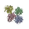
- PDB-5awg: Crystal structure of Hg-bound SufB-SufC-SufD complex from Escheri... -
+ Open data

- Basic information
| Entry | Database: PDB / ID: 5awg | ||||||
|---|---|---|---|---|---|---|---|
| Title | Crystal structure of Hg-bound SufB-SufC-SufD complex from Escherichia coli | ||||||
 Components Components |
| ||||||
 Keywords Keywords | TRANSPORT PROTEIN/PROTEIN BINDING /  Iron-Sulfur Clusters / Iron-Sulfur Proteins / ABC proteins / ABC ATPase / TRANSPORT PROTEIN-PROTEIN BINDING complex Iron-Sulfur Clusters / Iron-Sulfur Proteins / ABC proteins / ABC ATPase / TRANSPORT PROTEIN-PROTEIN BINDING complex | ||||||
| Function / homology |  Function and homology information Function and homology informationiron-sulfur cluster assembly complex / iron-sulfur cluster assembly / response to radiation / 2 iron, 2 sulfur cluster binding / 4 iron, 4 sulfur cluster binding / response to oxidative stress / ATP hydrolysis activity / ATP binding / cytosolSimilarity search - Function | ||||||
| Biological species |  Escherichia coli (E. coli) Escherichia coli (E. coli) | ||||||
| Method | X-RAY DIFFRACTION / SYNCHROTRON / MIR / Resolution: 4.278 Å | ||||||
 Authors Authors | Hirabayashi, K. / Wada, K. | ||||||
 Citation Citation | Journal: J Biol Chem / Year: 2015 Title: Functional Dynamics Revealed by the Structure of the SufBCD Complex, a Novel ATP-binding Cassette (ABC) Protein That Serves as a Scaffold for Iron-Sulfur Cluster Biogenesis. Authors: Kei Hirabayashi / Eiki Yuda / Naoyuki Tanaka / Sumie Katayama / Kenji Iwasaki / Takashi Matsumoto / Genji Kurisu / F Wayne Outten / Keiichi Fukuyama / Yasuhiro Takahashi / Kei Wada /   Abstract: ATP-binding cassette (ABC)-type ATPases are chemomechanical engines involved in diverse biological pathways. Recent genomic information reveals that ABC ATPase domains/subunits act not only in ABC ...ATP-binding cassette (ABC)-type ATPases are chemomechanical engines involved in diverse biological pathways. Recent genomic information reveals that ABC ATPase domains/subunits act not only in ABC transporters and structural maintenance of chromosome proteins, but also in iron-sulfur (Fe-S) cluster biogenesis. A novel type of ABC protein, the SufBCD complex, functions in the biosynthesis of nascent Fe-S clusters in almost all Eubacteria and Archaea, as well as eukaryotic chloroplasts. In this study, we determined the first crystal structure of the Escherichia coli SufBCD complex, which exhibits the common architecture of ABC proteins: two ABC ATPase components (SufC) with function-specific components (SufB-SufD protomers). Biochemical and physiological analyses based on this structure provided critical insights into Fe-S cluster assembly and revealed a dynamic conformational change driven by ABC ATPase activity. We propose a molecular mechanism for the biogenesis of the Fe-S cluster in the SufBCD complex. | ||||||
| History |
|
- Structure visualization
| Structure viewer | Molecule: MolmilJmol/JSmol |
|---|
- Downloads & links
-Download
| PDBx/mmCIF format | 5awg.cif.gz | 474.3 KB | Display | PDBx/mmCIF format |
|---|---|---|---|---|
| PDB format | pdb5awg.ent.gz | 393.4 KB | Display | PDB format |
| PDBx/mmJSON format | 5awg.json.gz | Tree view | PDBx/mmJSON format | |
| Others |  Other downloads Other downloads |
-Validation report
| Arichive directory | https://data.pdbj.org/pub/pdb/validation_reports/aw/5awgftp://data.pdbj.org/pub/pdb/validation_reports/aw/5awg | HTTPS FTP |
|---|
-Related structure data


-Links
 PDBj
PDBj


- Assembly
| Deposited unit | 
| ||||||||
|---|---|---|---|---|---|---|---|---|---|
| 1 | 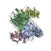
| ||||||||
| 2 | 
| ||||||||
| Unit cell |
|
-Components
| #1: Protein | Mass: 54800.070 Da / Num. of mol.: 2 Source method: isolated from a genetically manipulated source Source: (gene. exp.) Escherichia coli (strain K12) (bacteria)Strain: K12 / Gene: sufB, ynhE, b1683, JW5273 / Production host: Escherichia coli (E. coli) / References: UniProt: P77522#2: Protein | Mass: 46884.641 Da / Num. of mol.: 2 Source method: isolated from a genetically manipulated source Source: (gene. exp.) Escherichia coli (strain K12) (bacteria)Strain: K12 / Gene: sufD, ynhC, b1681, JW1671 / Production host: Escherichia coli (E. coli) / References: UniProt: P77689#3: Protein | Mass: 27613.332 Da / Num. of mol.: 4 Source method: isolated from a genetically manipulated source Source: (gene. exp.) Escherichia coli (strain K12) (bacteria)Strain: K12 / Gene: sufC, ynhD, b1682, JW1672 / Production host: Escherichia coli (E. coli) / References: UniProt: P77499#4: Chemical | ChemComp-HG / Mercury (element)  Mass: 200.590 Da / Num. of mol.: 4 / Source method: obtained synthetically / Formula: Hg Mass: 200.590 Da / Num. of mol.: 4 / Source method: obtained synthetically / Formula: Hg |
|---|
-Experimental details
-Experiment
| Experiment | Method: X-RAY DIFFRACTION |
|---|
- Sample preparation
| Crystal | Density Matthews: 3.04 Å3/Da / Density % sol: 59.48 % Description: THE ENTRY CONTAINS FRIEDEL PAIRS IN F_PLUS/MINUS COLUMNS. |
|---|---|
| Crystal grow | Temperature: 277 K / Method: vapor diffusion, sitting drop Details: pentaerythritol propoxylate (5/4 PO/OH), sodium citrate, potassium chloride |
-Data collection
| Diffraction | Mean temperature: 100 K |
|---|---|
| Diffraction source | Source: SYNCHROTRON / Site: SPring-8 / Beamline: BL44XU / Wavelength: 0.95 Å |
| Detector | Type: RAYONIX MX300HE / Detector: CCD / Date: Jun 28, 2014 |
| Radiation | Monochromator: Si(111) / Protocol: SINGLE WAVELENGTH / Monochromatic (M) / Laue (L): M / Scattering type: x-ray |
| Radiation wavelength | Wavelength: 0.95 Å / Relative weight: 1 |
| Reflection | Resolution: 4.278→50 Å / Num. obs: 50117 / % possible obs: 99.3 % / Redundancy: 5.6 % / Rmerge(I) obs: 0.127 / Net I/σ(I): 6.6 |
| Reflection shell | Resolution: 4.3→4.45 Å / Redundancy: 5.6 % / Rmerge(I) obs: 0.295 / Mean I/σ(I) obs: 5.2 / % possible all: 99.9 |
- Processing
| Software |
| |||||||||||||||||||||||||||||||||||||||||||||||||||||||||||||||||||||||||||||||||||||||||||||||||||||||||||||||||||||||||||||||||||||
|---|---|---|---|---|---|---|---|---|---|---|---|---|---|---|---|---|---|---|---|---|---|---|---|---|---|---|---|---|---|---|---|---|---|---|---|---|---|---|---|---|---|---|---|---|---|---|---|---|---|---|---|---|---|---|---|---|---|---|---|---|---|---|---|---|---|---|---|---|---|---|---|---|---|---|---|---|---|---|---|---|---|---|---|---|---|---|---|---|---|---|---|---|---|---|---|---|---|---|---|---|---|---|---|---|---|---|---|---|---|---|---|---|---|---|---|---|---|---|---|---|---|---|---|---|---|---|---|---|---|---|---|---|---|---|
| Refinement | Method to determine structure: MIR / Resolution: 4.278→43.942 Å / SU ML: 0.63 / Cross valid method: THROUGHOUT / σ(F): 1.35 / Phase error: 37.87 / Stereochemistry target values: ML Details: SF FILE CONTAINS FRIEDEL PAIRS UNDER I/F_MINUS AND I/F_PLUS COLUMNS.
| |||||||||||||||||||||||||||||||||||||||||||||||||||||||||||||||||||||||||||||||||||||||||||||||||||||||||||||||||||||||||||||||||||||
| Solvent computation | Shrinkage radii: 0.9 Å / VDW probe radii: 1.11 Å / Solvent model: FLAT BULK SOLVENT MODEL | |||||||||||||||||||||||||||||||||||||||||||||||||||||||||||||||||||||||||||||||||||||||||||||||||||||||||||||||||||||||||||||||||||||
| Refinement step | Cycle: LAST / Resolution: 4.278→43.942 Å
| |||||||||||||||||||||||||||||||||||||||||||||||||||||||||||||||||||||||||||||||||||||||||||||||||||||||||||||||||||||||||||||||||||||
| Refine LS restraints |
| |||||||||||||||||||||||||||||||||||||||||||||||||||||||||||||||||||||||||||||||||||||||||||||||||||||||||||||||||||||||||||||||||||||
| LS refinement shell |
|
