aspartate-semialdehyde dehydrogenase / aspartate-semialdehyde dehydrogenase activity / threonine biosynthetic process / lysine biosynthetic process via diaminopimelate / NAD binding / NADP binding / protein dimerization activity Similarity search - Function
Mass: 18.015 Da / Num. of mol.: 543 / Source method: isolated from a natural source / Formula: H2O
Sequence details
THE SEQUENCE OF THIS PROTEIN WAS NOT AVAILABLE AT THE UNIPROT KNOWLEDGEBASE DATABASE (UNIPROTKB) AT ...THE SEQUENCE OF THIS PROTEIN WAS NOT AVAILABLE AT THE UNIPROT KNOWLEDGEBASE DATABASE (UNIPROTKB) AT THE TIME OF DEPOSITION. N-TERMINAL RESIDUES MGSSHHHHHHSSGLVPRGSHM ARE THE EXPRESSION TAGS.
-
Experimental details
-
Experiment
Experiment
Method: X-RAY DIFFRACTION / Number of used crystals: 1
-
Sample preparation
Crystal
Density Matthews: 2.75 Å3/Da / Density % sol: 55.35 %
Resolution: 2.553→46.133 Å / SU ML: 0.41 / Cross valid method: FREE R-VALUE / σ(F): 1.35 / Phase error: 28.16 / Stereochemistry target values: ML Details: DURING DATA COLLECTION, F+ AND F- WERE CONSIDERED AS DIFFERENT REFLECTION BECAUSE THE FRIEDEL'S LAW WAS FALSE. AFTER MOLECULAR REPLACEMENT FOR STRUCTURE DETERMINATION, IN THE REFINEMENT F+ ...Details: DURING DATA COLLECTION, F+ AND F- WERE CONSIDERED AS DIFFERENT REFLECTION BECAUSE THE FRIEDEL'S LAW WAS FALSE. AFTER MOLECULAR REPLACEMENT FOR STRUCTURE DETERMINATION, IN THE REFINEMENT F+ AND F- WERE MERGED THEREFORE THE REFLECTION COUNTED WERE AROUND 50% OF DATA COLLECTION.
Rfactor
Num. reflection
% reflection
Rfree
0.2725
4385
5.01 %
Rwork
0.2267
-
-
obs
0.229
87486
99.73 %
Solvent computation
Shrinkage radii: 0.98 Å / VDW probe radii: 1.2 Å / Solvent model: FLAT BULK SOLVENT MODEL / Bsol: 29.802 Å2 / ksol: 0.315 e/Å3
Displacement parameters
Baniso -1
Baniso -2
Baniso -3
1-
0.3413 Å2
0 Å2
-0 Å2
2-
-
0.3413 Å2
-0 Å2
3-
-
-
-0.6827 Å2
Refinement step
Cycle: LAST / Resolution: 2.553→46.133 Å
Protein
Nucleic acid
Ligand
Solvent
Total
Num. atoms
16043
0
126
543
16712
Refine LS restraints
Refine-ID
Type
Dev ideal
Number
X-RAY DIFFRACTION
f_bond_d
0.004
16604
X-RAY DIFFRACTION
f_angle_d
1.056
22558
X-RAY DIFFRACTION
f_dihedral_angle_d
13.081
6171
X-RAY DIFFRACTION
f_chiral_restr
0.076
2580
X-RAY DIFFRACTION
f_plane_restr
0.004
2953
LS refinement shell
Resolution (Å)
Rfactor Rfree
Num. reflection Rfree
Rfactor Rwork
Num. reflection Rwork
Refine-ID
% reflection obs (%)
2.5527-2.5817
0.4106
124
0.3073
2760
X-RAY DIFFRACTION
99
2.5817-2.6121
0.3018
130
0.2943
2719
X-RAY DIFFRACTION
100
2.6121-2.6439
0.4132
133
0.2923
2742
X-RAY DIFFRACTION
100
2.6439-2.6774
0.338
137
0.2904
2745
X-RAY DIFFRACTION
100
2.6774-2.7126
0.4121
147
0.2947
2751
X-RAY DIFFRACTION
100
2.7126-2.7498
0.374
152
0.2897
2735
X-RAY DIFFRACTION
100
2.7498-2.7891
0.3539
136
0.2819
2745
X-RAY DIFFRACTION
100
2.7891-2.8307
0.3223
134
0.2653
2746
X-RAY DIFFRACTION
100
2.8307-2.8749
0.3503
142
0.277
2780
X-RAY DIFFRACTION
100
2.8749-2.922
0.3371
173
0.2792
2711
X-RAY DIFFRACTION
100
2.922-2.9724
0.3086
143
0.2725
2767
X-RAY DIFFRACTION
100
2.9724-3.0264
0.3554
152
0.2923
2757
X-RAY DIFFRACTION
100
3.0264-3.0846
0.3189
143
0.2697
2753
X-RAY DIFFRACTION
100
3.0846-3.1476
0.3007
146
0.2629
2751
X-RAY DIFFRACTION
100
3.1476-3.216
0.3177
148
0.2607
2760
X-RAY DIFFRACTION
100
3.216-3.2908
0.2824
154
0.2519
2716
X-RAY DIFFRACTION
100
3.2908-3.3731
0.2971
158
0.2464
2766
X-RAY DIFFRACTION
100
3.3731-3.4643
0.2886
127
0.236
2794
X-RAY DIFFRACTION
100
3.4643-3.5662
0.2844
157
0.2278
2744
X-RAY DIFFRACTION
100
3.5662-3.6812
0.2425
141
0.2259
2791
X-RAY DIFFRACTION
100
3.6812-3.8127
0.2462
156
0.2243
2768
X-RAY DIFFRACTION
100
3.8127-3.9653
0.2666
138
0.2082
2768
X-RAY DIFFRACTION
100
3.9653-4.1457
0.213
160
0.2009
2798
X-RAY DIFFRACTION
100
4.1457-4.3641
0.2171
148
0.1809
2765
X-RAY DIFFRACTION
100
4.3641-4.6373
0.2343
140
0.1784
2810
X-RAY DIFFRACTION
100
4.6373-4.9949
0.2234
143
0.1737
2789
X-RAY DIFFRACTION
100
4.9949-5.4968
0.2454
148
0.1916
2812
X-RAY DIFFRACTION
100
5.4968-6.2905
0.2892
153
0.2222
2838
X-RAY DIFFRACTION
100
6.2905-7.9189
0.2458
171
0.2032
2833
X-RAY DIFFRACTION
100
7.9189-46.1402
0.2125
151
0.1968
2887
X-RAY DIFFRACTION
97
+
About Yorodumi
-
News
-
Feb 9, 2022. New format data for meta-information of EMDB entries
New format data for meta-information of EMDB entries
Version 3 of the EMDB header file is now the official format.
The previous official version 1.9 will be removed from the archive.
In the structure databanks used in Yorodumi, some data are registered as the other names, "COVID-19 virus" and "2019-nCoV". Here are the details of the virus and the list of structure data.
Jan 31, 2019. EMDB accession codes are about to change! (news from PDBe EMDB page)
EMDB accession codes are about to change! (news from PDBe EMDB page)
The allocation of 4 digits for EMDB accession codes will soon come to an end. Whilst these codes will remain in use, new EMDB accession codes will include an additional digit and will expand incrementally as the available range of codes is exhausted. The current 4-digit format prefixed with “EMD-” (i.e. EMD-XXXX) will advance to a 5-digit format (i.e. EMD-XXXXX), and so on. It is currently estimated that the 4-digit codes will be depleted around Spring 2019, at which point the 5-digit format will come into force.
The EM Navigator/Yorodumi systems omit the EMD- prefix.
Related info.:Q: What is EMD? / ID/Accession-code notation in Yorodumi/EM Navigator
Yorodumi is a browser for structure data from EMDB, PDB, SASBDB, etc.
This page is also the successor to EM Navigator detail page, and also detail information page/front-end page for Omokage search.
The word "yorodu" (or yorozu) is an old Japanese word meaning "ten thousand". "mi" (miru) is to see.
Related info.:EMDB / PDB / SASBDB / Comparison of 3 databanks / Yorodumi Search / Aug 31, 2016. New EM Navigator & Yorodumi / Yorodumi Papers / Jmol/JSmol / Function and homology information / Changes in new EM Navigator and Yorodumi
 Movie
Movie Controller

 Yorodumi
 Open data
 Basic information
 Components
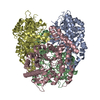
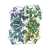
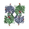
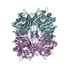
 Aspartate-semialdehyde dehydrogenase
  Keywords
 Function and homology information
 Authors
 Citation
 Structure visualization
 Downloads & links
 Other downloads


 PDBj
PDBj Assembly


 Mass: 96.063 Da / Num. of mol.: 6 / Source method: obtained synthetically / Formula: SO4
 Mass: 743.405 Da / Num. of mol.: 2 / Source method: obtained synthetically / Formula: C21H28N7O17P3
 Mass: 18.015 Da / Num. of mol.: 543 / Source method: isolated from a natural source / Formula: H2O
 Sample preparation
 / Beamline: BL17U / Wavelength: 0.97876 Å
 Processing