 Movie
Movie Controller

[English] 日本語
 Yorodumi
- PDB-4zc1: Crystal Structure of type II Dehydroquinate dehydratase from Acin... -
+ Open data

- Basic information
| Entry | Database: PDB / ID: 4zc1 | ||||||
|---|---|---|---|---|---|---|---|
| Title | Crystal Structure of type II Dehydroquinate dehydratase from Acinetobacter baumannii with a different crystal form at 2.52 A Resolution | ||||||
 Components Components | 3-dehydroquinate dehydratase | ||||||
 Keywords Keywords | LYASE / 3-DEHYDROQUINASE / TYPE II DHQASE | ||||||
| Function / homology |  Function and homology information3-dehydroquinate dehydratase / 3-dehydroquinate dehydratase activity / chorismate biosynthetic process / aromatic amino acid family biosynthetic process / amino acid biosynthetic process Function and homology information3-dehydroquinate dehydratase / 3-dehydroquinate dehydratase activity / chorismate biosynthetic process / aromatic amino acid family biosynthetic process / amino acid biosynthetic processSimilarity search - Function | ||||||
| Biological species |  Acinetobacter baumannii (bacteria) Acinetobacter baumannii (bacteria) | ||||||
| Method | X-RAY DIFFRACTION / SYNCHROTRON / MOLECULAR REPLACEMENT / Resolution: 2.52 Å | ||||||
 Authors Authors | Iqbal, N. / Kumar, M. / Kaur, P. / Sharma, S. / Singh, T.P. | ||||||
 Citation Citation | Journal: To Be Published Title: Crystal Structure of type II Dehydroquinate dehydratase from Acinetobacter baumannii with different crystal form at 2.52 A Resolution Authors: Iqbal, N. / Kumar, M. / Kaur, P. / Sharma, S. / Singh, T.P. | ||||||
| History |
|
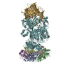
- Structure visualization
| Structure viewer | Molecule: MolmilJmol/JSmol |
|---|
- Downloads & links
-Download
| PDBx/mmCIF format | 4zc1.cif.gz | 340.3 KB | Display | PDBx/mmCIF format |
|---|---|---|---|---|
| PDB format | pdb4zc1.ent.gz | 280 KB | Display | PDB format |
| PDBx/mmJSON format | 4zc1.json.gz | Tree view | PDBx/mmJSON format | |
| Others |  Other downloads Other downloads |
-Validation report
| Arichive directory | https://data.pdbj.org/pub/pdb/validation_reports/zc/4zc1ftp://data.pdbj.org/pub/pdb/validation_reports/zc/4zc1 | HTTPS FTP |
|---|
-Related structure data
| Related structure data |  4rc9S S: Starting model for refinement |
|---|---|
| Similar structure data |


-Links
 PDBj
PDBj
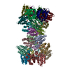
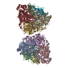
- Assembly
| Deposited unit | 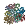
| ||||||||
|---|---|---|---|---|---|---|---|---|---|
| 1 | 
| ||||||||
| Unit cell |
|
-Components
| #1: Protein | / 3-dehydroquinase / Type II DHQase Mass: 16007.288 Da / Num. of mol.: 12 Source method: isolated from a genetically manipulated source Source: (gene. exp.) Acinetobacter baumannii (strain ATCC 17978 / NCDC KC 755) (bacteria)Strain: ATCC 17978 / NCDC KC 755 / Gene: aroQ, A1S_2009 / Plasmid: Pet28a / Production host: Escherichia coli (E. coli) / References: UniProt: A3M692, 3-dehydroquinate dehydratase#2: Water | ChemComp-HOH / | Water Mass: 18.015 Da / Num. of mol.: 393 / Source method: isolated from a natural source / Formula: H2O Mass: 18.015 Da / Num. of mol.: 393 / Source method: isolated from a natural source / Formula: H2O |
|---|
-Experimental details
-Experiment
| Experiment | Method: X-RAY DIFFRACTION |
|---|
- Sample preparation
| Crystal | Density Matthews: 2.46 Å3/Da / Density % sol: 49.95 % Description: THE ENTRY CONTAINS FRIEDEL PAIRS IN F_PLUS/MINUS COLUMNS. |
|---|---|
| Crystal grow | Temperature: 298 K / Method: vapor diffusion, hanging drop / pH: 8 / Details: 30% PEG3350, MgSO4, Tris buffer / PH range: 8 |
-Data collection
| Diffraction | Mean temperature: 77 K / Ambient temp details: CCD |
|---|---|
| Diffraction source | Source: SYNCHROTRON / Site: ESRF  / Beamline: BM14 / Wavelength: 0.97 Å / Beamline: BM14 / Wavelength: 0.97 Å |
| Detector | Type: MARRESEARCH / Detector: CCD / Date: Nov 14, 2014 |
| Radiation | Monochromator: Graphite / Protocol: SINGLE WAVELENGTH / Monochromatic (M) / Laue (L): M / Scattering type: x-ray |
| Radiation wavelength | Wavelength: 0.97 Å / Relative weight: 1 |
| Reflection | Resolution: 2.52→50 Å / Num. all: 248902 / Num. obs: 62069 / % possible obs: 100 % / Redundancy: 4 % / Rsym value: 0.1 / Net I/σ(I): 13.3 |
| Reflection shell | Resolution: 2.52→2.56 Å / Redundancy: 4 % / Rmerge(I) obs: 0.639 / Mean I/σ(I) obs: 2 / % possible all: 100 |
- Processing
| Software |
| ||||||||||||||||||||||||||||||||||||||||||||||||||||||||||||||||||||||||||||||||||||||||||||||||||||||||||||||||||||||||||||||||||||||||||||||||||||||||||||||||||||||||||||||||||||||
|---|---|---|---|---|---|---|---|---|---|---|---|---|---|---|---|---|---|---|---|---|---|---|---|---|---|---|---|---|---|---|---|---|---|---|---|---|---|---|---|---|---|---|---|---|---|---|---|---|---|---|---|---|---|---|---|---|---|---|---|---|---|---|---|---|---|---|---|---|---|---|---|---|---|---|---|---|---|---|---|---|---|---|---|---|---|---|---|---|---|---|---|---|---|---|---|---|---|---|---|---|---|---|---|---|---|---|---|---|---|---|---|---|---|---|---|---|---|---|---|---|---|---|---|---|---|---|---|---|---|---|---|---|---|---|---|---|---|---|---|---|---|---|---|---|---|---|---|---|---|---|---|---|---|---|---|---|---|---|---|---|---|---|---|---|---|---|---|---|---|---|---|---|---|---|---|---|---|---|---|---|---|---|---|
| Refinement | Method to determine structure: MOLECULAR REPLACEMENT Starting model: 4RC9 Resolution: 2.52→50 Å / Cor.coef. Fo:Fc: 0.917 / Cor.coef. Fo:Fc free: 0.859 / SU B: 13.157 / SU ML: 0.286 / Data cutoff high absF: 0 / Data cutoff low absF: 0 / Cross valid method: THROUGHOUT / σ(F): 0 / ESU R: 1.15 / ESU R Free: 0.342 / Stereochemistry target values: MAXIMUM LIKELIHOOD Details: HYDROGENS HAVE BEEN ADDED IN THE RIDING POSITIONS, SF FILE CONTAINS FRIEDEL PAIRS UNDER I/F_MINUS AND I/F_PLUS COLUMNS.
| ||||||||||||||||||||||||||||||||||||||||||||||||||||||||||||||||||||||||||||||||||||||||||||||||||||||||||||||||||||||||||||||||||||||||||||||||||||||||||||||||||||||||||||||||||||||
| Solvent computation | Ion probe radii: 0.8 Å / Shrinkage radii: 0.8 Å / VDW probe radii: 1.2 Å / Solvent model: MASK | ||||||||||||||||||||||||||||||||||||||||||||||||||||||||||||||||||||||||||||||||||||||||||||||||||||||||||||||||||||||||||||||||||||||||||||||||||||||||||||||||||||||||||||||||||||||
| Displacement parameters | Biso mean: 39.165 Å2
| ||||||||||||||||||||||||||||||||||||||||||||||||||||||||||||||||||||||||||||||||||||||||||||||||||||||||||||||||||||||||||||||||||||||||||||||||||||||||||||||||||||||||||||||||||||||
| Refinement step | Cycle: 1 / Resolution: 2.52→50 Å
| ||||||||||||||||||||||||||||||||||||||||||||||||||||||||||||||||||||||||||||||||||||||||||||||||||||||||||||||||||||||||||||||||||||||||||||||||||||||||||||||||||||||||||||||||||||||
| Refine LS restraints |
|