 Movie
Movie Controller

[English] 日本語
 Yorodumi
- PDB-4z6w: Structure of H200N variant of Homoprotocatechuate 2,3-Dioxygenase... -
+ Open data

- Basic information
| Entry | Database: PDB / ID: 4z6w | |||||||||
|---|---|---|---|---|---|---|---|---|---|---|
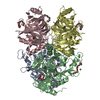
| Title | Structure of H200N variant of Homoprotocatechuate 2,3-Dioxygenase from B.fuscum in complex with 4-nitrocatechol at 1.57 Ang resolution | |||||||||
 Components Components | Homoprotocatechuate 2,3-dioxygenase | |||||||||
 Keywords Keywords |  OXIDOREDUCTASE / Dioxygenase / 2-His-1-carboxylate facial triad / oxygen activation / acid-base catalysis OXIDOREDUCTASE / Dioxygenase / 2-His-1-carboxylate facial triad / oxygen activation / acid-base catalysis | |||||||||
| Function / homology |  Function and homology information Function and homology information | |||||||||
| Biological species |  Brevibacterium fuscum (bacteria) Brevibacterium fuscum (bacteria) | |||||||||
| Method | X-RAY DIFFRACTION / SYNCHROTRON / MOLECULAR REPLACEMENT / Resolution: 1.57 Å | |||||||||
 Authors Authors | Kovaleva, E.G. / Lipscomb, J.D. | |||||||||
| Funding support |  United Kingdom, United Kingdom,  United States, 2items United States, 2items
| |||||||||
 Citation Citation | Journal: Biochemistry / Year: 2015 Title: Structural Basis for Substrate and Oxygen Activation in Homoprotocatechuate 2,3-Dioxygenase: Roles of Conserved Active Site Histidine 200. Authors: Kovaleva, E.G. / Rogers, M.S. / Lipscomb, J.D. | |||||||||
| History |
|
- Structure visualization
| Structure viewer | Molecule: MolmilJmol/JSmol |
|---|
- Downloads & links
-Download
| PDBx/mmCIF format | 4z6w.cif.gz | 621.4 KB | Display | PDBx/mmCIF format |
|---|---|---|---|---|
| PDB format | pdb4z6w.ent.gz | 510.4 KB | Display | PDB format |
| PDBx/mmJSON format | 4z6w.json.gz | Tree view | PDBx/mmJSON format | |
| Others |  Other downloads Other downloads |
-Validation report
| Arichive directory | https://data.pdbj.org/pub/pdb/validation_reports/z6/4z6wftp://data.pdbj.org/pub/pdb/validation_reports/z6/4z6w | HTTPS FTP |
|---|
-Related structure data
| Related structure data |  4z6lC  4z6mC  4z6nC  4z6oC  4z6pC  4z6qC  4z6rC  4z6sC  4z6tC  4z6uC  4z6vC  4z6zC  3ojtS C: citing same article ( S: Starting model for refinement |
|---|---|
| Similar structure data |


-Links
 PDBj
PDBj


- Assembly
| Deposited unit | 
| |||||||||
|---|---|---|---|---|---|---|---|---|---|---|
| 1 |
| |||||||||
| Unit cell |
| |||||||||
| Components on special symmetry positions |
|
-Components
-Protein , 1 types, 4 molecules ABCD
| #1: Protein | Mass: 41731.277 Da / Num. of mol.: 4 / Mutation: H200N Source method: isolated from a genetically manipulated source Source: (gene. exp.) Brevibacterium fuscum (bacteria) / Plasmid: pYZW204 / Production host: Escherichia coli (E. coli) / Strain (production host): Jm109 / References: UniProt: Q45135 |
|---|
-Non-polymers , 7 types, 1731 molecules 


| #2: Chemical | ChemComp-FE2 /  Mass: 55.845 Da / Num. of mol.: 4 / Source method: obtained synthetically / Formula: Fe Mass: 55.845 Da / Num. of mol.: 4 / Source method: obtained synthetically / Formula: Fe#3: Chemical | ChemComp-P6G / Polyethylene glycol Mass: 282.331 Da / Num. of mol.: 5 / Source method: obtained synthetically / Formula: C12H26O7 / Comment: precipitant*YM Mass: 282.331 Da / Num. of mol.: 5 / Source method: obtained synthetically / Formula: C12H26O7 / Comment: precipitant*YM#4: Chemical | ChemComp-4NC /  Mass: 155.108 Da / Num. of mol.: 4 / Source method: obtained synthetically / Formula: C6H5NO4 Mass: 155.108 Da / Num. of mol.: 4 / Source method: obtained synthetically / Formula: C6H5NO4#5: Chemical | Chloride Mass: 35.453 Da / Num. of mol.: 3 / Source method: obtained synthetically / Formula: Cl Mass: 35.453 Da / Num. of mol.: 3 / Source method: obtained synthetically / Formula: Cl#6: Chemical | ChemComp-CA / |  Mass: 40.078 Da / Num. of mol.: 1 / Source method: obtained synthetically / Formula: Ca Mass: 40.078 Da / Num. of mol.: 1 / Source method: obtained synthetically / Formula: Ca#7: Chemical | Diethylene glycol Mass: 106.120 Da / Num. of mol.: 2 / Source method: obtained synthetically / Formula: C4H10O3 Mass: 106.120 Da / Num. of mol.: 2 / Source method: obtained synthetically / Formula: C4H10O3#8: Water | ChemComp-HOH / | WaterMass: 18.015 Da / Num. of mol.: 1712 / Source method: isolated from a natural source / Formula: H2O |
|---|
-Experimental details
-Experiment
| Experiment | Method: X-RAY DIFFRACTION / Number of used crystals: 1 |
|---|
- Sample preparation
| Crystal | Density Matthews: 2.4 Å3/Da / Density % sol: 48.68 % |
|---|---|
| Crystal grow | Temperature: 293 K / Method: vapor diffusion, hanging drop / pH: 7 / Details: 14% PEG6000, 0.1M calcium acetate, 0.1M MOPS |
-Data collection
| Diffraction | Mean temperature: 100 K | ||||||||||||||||||||||||||||||||||||||||||||||||||||||||||||||||||||||||||||||||||||||||||||||||||||||||||||||
|---|---|---|---|---|---|---|---|---|---|---|---|---|---|---|---|---|---|---|---|---|---|---|---|---|---|---|---|---|---|---|---|---|---|---|---|---|---|---|---|---|---|---|---|---|---|---|---|---|---|---|---|---|---|---|---|---|---|---|---|---|---|---|---|---|---|---|---|---|---|---|---|---|---|---|---|---|---|---|---|---|---|---|---|---|---|---|---|---|---|---|---|---|---|---|---|---|---|---|---|---|---|---|---|---|---|---|---|---|---|---|---|
| Diffraction source | Source: SYNCHROTRON / Site: SOLEIL  / Beamline: PROXIMA 1 / Wavelength: 0.9801 Å / Beamline: PROXIMA 1 / Wavelength: 0.9801 Å | ||||||||||||||||||||||||||||||||||||||||||||||||||||||||||||||||||||||||||||||||||||||||||||||||||||||||||||||
| Detector | Type: DECTRIS PILATUS 6M / Detector: PIXEL / Date: Apr 1, 2012 | ||||||||||||||||||||||||||||||||||||||||||||||||||||||||||||||||||||||||||||||||||||||||||||||||||||||||||||||
| Radiation | Protocol: SINGLE WAVELENGTH / Monochromatic (M) / Laue (L): M / Scattering type: x-ray | ||||||||||||||||||||||||||||||||||||||||||||||||||||||||||||||||||||||||||||||||||||||||||||||||||||||||||||||
| Radiation wavelength | Wavelength: 0.9801 Å / Relative weight: 1 | ||||||||||||||||||||||||||||||||||||||||||||||||||||||||||||||||||||||||||||||||||||||||||||||||||||||||||||||
| Reflection | Resolution: 1.57→48 Å / Num. all: 218224 / Num. obs: 218224 / % possible obs: 98.1 % / Redundancy: 6.7 % / Rpim(I) all: 0.037 / Rrim(I) all: 0.097 / Rsym value: 0.09 / Net I/av σ(I): 6.3 / Net I/σ(I): 12.6 / Num. measured all: 1452866 | ||||||||||||||||||||||||||||||||||||||||||||||||||||||||||||||||||||||||||||||||||||||||||||||||||||||||||||||
| Reflection shell | Diffraction-ID: 1 / Rejects: 0
|
- Processing
| Software |
| |||||||||||||||||||||||||||||||||||||||||||||||||||||||||||||||||||||||||||||||||||||||||||||||||||||||||||||||||||||||||||||
|---|---|---|---|---|---|---|---|---|---|---|---|---|---|---|---|---|---|---|---|---|---|---|---|---|---|---|---|---|---|---|---|---|---|---|---|---|---|---|---|---|---|---|---|---|---|---|---|---|---|---|---|---|---|---|---|---|---|---|---|---|---|---|---|---|---|---|---|---|---|---|---|---|---|---|---|---|---|---|---|---|---|---|---|---|---|---|---|---|---|---|---|---|---|---|---|---|---|---|---|---|---|---|---|---|---|---|---|---|---|---|---|---|---|---|---|---|---|---|---|---|---|---|---|---|---|---|
| Refinement | Method to determine structure: MOLECULAR REPLACEMENT Starting model: 3OJT Resolution: 1.57→45.73 Å / Cor.coef. Fo:Fc: 0.978 / Cor.coef. Fo:Fc free: 0.97 / WRfactor Rfree: 0.155 / WRfactor Rwork: 0.1291 / FOM work R set: 0.8953 / SU B: 2.826 / SU ML: 0.05 / SU R Cruickshank DPI: 0.0665 / SU Rfree: 0.0675 / Cross valid method: THROUGHOUT / σ(F): 0 / ESU R: 0.066 / ESU R Free: 0.068 / Stereochemistry target values: MAXIMUM LIKELIHOOD Details: HYDROGENS HAVE BEEN ADDED IN THE RIDING POSITIONS U VALUES : WITH TLS ADDED
| |||||||||||||||||||||||||||||||||||||||||||||||||||||||||||||||||||||||||||||||||||||||||||||||||||||||||||||||||||||||||||||
| Solvent computation | Ion probe radii: 0.8 Å / Shrinkage radii: 0.8 Å / VDW probe radii: 1.2 Å / Solvent model: BABINET MODEL WITH MASK | |||||||||||||||||||||||||||||||||||||||||||||||||||||||||||||||||||||||||||||||||||||||||||||||||||||||||||||||||||||||||||||
| Displacement parameters | Biso max: 76.52 Å2 / Biso mean: 20.802 Å2 / Biso min: 8.74 Å2
| |||||||||||||||||||||||||||||||||||||||||||||||||||||||||||||||||||||||||||||||||||||||||||||||||||||||||||||||||||||||||||||
| Refinement step | Cycle: final / Resolution: 1.57→45.73 Å
| |||||||||||||||||||||||||||||||||||||||||||||||||||||||||||||||||||||||||||||||||||||||||||||||||||||||||||||||||||||||||||||
| Refine LS restraints |
| |||||||||||||||||||||||||||||||||||||||||||||||||||||||||||||||||||||||||||||||||||||||||||||||||||||||||||||||||||||||||||||
| LS refinement shell | Resolution: 1.57→1.611 Å / Total num. of bins used: 20
| |||||||||||||||||||||||||||||||||||||||||||||||||||||||||||||||||||||||||||||||||||||||||||||||||||||||||||||||||||||||||||||
| Refinement TLS params. | Method: refined / Refine-ID: X-RAY DIFFRACTION
| |||||||||||||||||||||||||||||||||||||||||||||||||||||||||||||||||||||||||||||||||||||||||||||||||||||||||||||||||||||||||||||
| Refinement TLS group |
|
