 Movie
Movie Controller


+ Open data

- Basic information
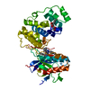
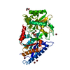
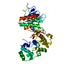
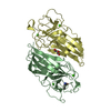
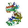
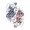
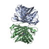
| Entry | Database: PDB / ID: 4yno | |||||||||
|---|---|---|---|---|---|---|---|---|---|---|
| Title | Crystal structure of MAPK13 at INACTIVE FORM | |||||||||
 Components Components | Mitogen-activated protein kinase 13 P38 mitogen-activated protein kinases P38 mitogen-activated protein kinases | |||||||||
 Keywords Keywords | TRANSFERASE / P38 KINASE | |||||||||
| Function / homology |  Function and homology information Function and homology informationcellular response to anisomycin / cellular response to sorbitol / cellular response to sodium arsenite / response to osmotic stress / DSCAM interactions / MAP kinase activity / mitogen-activated protein kinase / cellular response to interleukin-1 / stress-activated MAPK cascade / p38MAPK events ...cellular response to anisomycin / cellular response to sorbitol / cellular response to sodium arsenite / response to osmotic stress / DSCAM interactions / MAP kinase activity / mitogen-activated protein kinase / cellular response to interleukin-1 / stress-activated MAPK cascade / p38MAPK events / NOD1/2 Signaling Pathway / cellular response to hydrogen peroxide / VEGFA-VEGFR2 Pathway / positive regulation of inflammatory response / positive regulation of interleukin-6 production / cellular response to UV / peptidyl-serine phosphorylation / intracellular signal transduction / cell cycle / protein serine kinase activity / protein serine/threonine kinase activity / ATP binding / nucleus / cytosol / cytoplasmSimilarity search - Function | |||||||||
| Biological species |  Homo sapiens (human) Homo sapiens (human) | |||||||||
| Method | X-RAY DIFFRACTION / SYNCHROTRON / MOLECULAR REPLACEMENT / Resolution: 1.7 Å | |||||||||
 Authors Authors | Miller, C.A. / Brett, T.J. | |||||||||
| Funding support |  United States, 1items United States, 1items
| |||||||||
 Citation Citation | Journal: J.Clin.Invest. / Year: 2012 Title: IL-13-induced airway mucus production is attenuated by MAPK13 inhibition. Authors: Alevy, Y.G. / Patel, A.C. / Romero, A.G. / Patel, D.A. / Tucker, J. / Roswit, W.T. / Miller, C.A. / Heier, R.F. / Byers, D.E. / Brett, T.J. / Holtzman, M.J. #1: Journal: To Be PublishedTitle: The crystal structure of phosphorylated MAPK13 reveals common structural features and differences in p38 MAPK family activation Authors: Yurtsever, Z. / Scheaffer, S.M. / Romero, A.G. / Holtzman, M.J. / Brett, T.J. | |||||||||
| History |
|
- Structure visualization
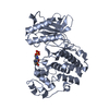
| Structure viewer | Molecule: MolmilJmol/JSmol |
|---|
- Downloads & links
-Download
| PDBx/mmCIF format | 4yno.cif.gz | 92.6 KB | Display | PDBx/mmCIF format |
|---|---|---|---|---|
| PDB format | pdb4yno.ent.gz | 67.1 KB | Display | PDB format |
| PDBx/mmJSON format | 4yno.json.gz | Tree view | PDBx/mmJSON format | |
| Others |  Other downloads Other downloads |
-Validation report
| Arichive directory | https://data.pdbj.org/pub/pdb/validation_reports/yn/4ynoftp://data.pdbj.org/pub/pdb/validation_reports/yn/4yno | HTTPS FTP |
|---|
-Related structure data
| Related structure data | 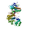 4eyjC 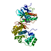 4eymC  3coiS C: citing same article ( S: Starting model for refinement |
|---|---|
| Similar structure data |

-Links
 PDBj
PDBj


- Assembly
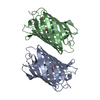
| Deposited unit | 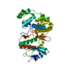
| ||||||||
|---|---|---|---|---|---|---|---|---|---|
| 1 |
| ||||||||
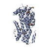
| Unit cell |
|
-Components
| #1: Protein | P38 mitogen-activated protein kinases / MAPK 13 / Mitogen-activated protein kinase p38 delta / MAP kinase p38 delta / Stress-activated ...MAPK 13 / Mitogen-activated protein kinase p38 delta / MAP kinase p38 delta / Stress-activated protein kinase 4 Mass: 42633.898 Da / Num. of mol.: 1 Source method: isolated from a genetically manipulated source Source: (gene. exp.) Homo sapiens (human) / Gene: MAPK13, PRKM13, SAPK4 / Plasmid: PET28A / Production host:  Escherichia coli (E. coli) / Strain (production host): ROSETTA2 (DE3) Escherichia coli (E. coli) / Strain (production host): ROSETTA2 (DE3)References: UniProt: O15264, mitogen-activated protein kinase |
|---|---|
| #2: Water | ChemComp-HOH / Water Mass: 18.015 Da / Num. of mol.: 315 / Source method: isolated from a natural source / Formula: H2O Mass: 18.015 Da / Num. of mol.: 315 / Source method: isolated from a natural source / Formula: H2O |
-Experimental details
-Experiment
| Experiment | Method: X-RAY DIFFRACTION |
|---|
- Sample preparation
| Crystal | Density Matthews: 2.29 Å3/Da / Density % sol: 46.37 % |
|---|---|
| Crystal grow | Temperature: 290 K / Method: vapor diffusion, hanging drop / pH: 6.5 / Details: 50 MM AMMONIUM TARTRATE, 18% PEG 3350 |
-Data collection
| Diffraction | Mean temperature: 105 K |
|---|---|
| Diffraction source | Source: SYNCHROTRON / Site: APS / Beamline: 19-ID / Wavelength: 1.007 Å |
| Detector | Type: ADSC QUANTUM 315 / Detector: CCD / Date: Aug 20, 2011 |
| Radiation | Monochromator: SI 111 / Protocol: SINGLE WAVELENGTH / Monochromatic (M) / Laue (L): M / Scattering type: x-ray |
| Radiation wavelength | Wavelength: 1.007 Å / Relative weight: 1 |
| Reflection | Resolution: 1.7→50 Å / Num. all: 43897 / Num. obs: 43897 / % possible obs: 99.3 % / Redundancy: 6.7 % / Rmerge(I) obs: 0.065 / Rsym value: 0.065 / Net I/σ(I): 29.6 |
| Reflection shell | Resolution: 1.7→1.76 Å / Redundancy: 6.9 % / Rmerge(I) obs: 0.504 / Mean I/σ(I) obs: 4.1 / % possible all: 99.7 |
- Processing
| Software |
| |||||||||||||||||||||||||||||||||||||||||||||||||||||||||||||||||||||||||||||||||||||||||||||||||||||||||||||||||||||||
|---|---|---|---|---|---|---|---|---|---|---|---|---|---|---|---|---|---|---|---|---|---|---|---|---|---|---|---|---|---|---|---|---|---|---|---|---|---|---|---|---|---|---|---|---|---|---|---|---|---|---|---|---|---|---|---|---|---|---|---|---|---|---|---|---|---|---|---|---|---|---|---|---|---|---|---|---|---|---|---|---|---|---|---|---|---|---|---|---|---|---|---|---|---|---|---|---|---|---|---|---|---|---|---|---|---|---|---|---|---|---|---|---|---|---|---|---|---|---|---|---|
| Refinement | Method to determine structure: MOLECULAR REPLACEMENT Starting model: PDB entry 3COI Resolution: 1.7→36.839 Å / SU ML: 0.16 / Cross valid method: FREE R-VALUE / σ(F): 1.34 / Phase error: 23.2 / Stereochemistry target values: ML
| |||||||||||||||||||||||||||||||||||||||||||||||||||||||||||||||||||||||||||||||||||||||||||||||||||||||||||||||||||||||
| Solvent computation | Shrinkage radii: 0.9 Å / VDW probe radii: 1.11 Å / Solvent model: FLAT BULK SOLVENT MODEL | |||||||||||||||||||||||||||||||||||||||||||||||||||||||||||||||||||||||||||||||||||||||||||||||||||||||||||||||||||||||
| Refinement step | Cycle: LAST / Resolution: 1.7→36.839 Å
| |||||||||||||||||||||||||||||||||||||||||||||||||||||||||||||||||||||||||||||||||||||||||||||||||||||||||||||||||||||||
| Refine LS restraints |
| |||||||||||||||||||||||||||||||||||||||||||||||||||||||||||||||||||||||||||||||||||||||||||||||||||||||||||||||||||||||
| LS refinement shell |
|
