 Movie
Movie Controller

[English] 日本語
 Yorodumi
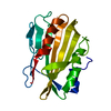
- PDB-4xcp: Fatty Acid and Retinol binding protein Na-FAR-1 from Necator amer... -
+ Open data

- Basic information
| Entry | Database: PDB / ID: 4xcp | ||||||||||||
|---|---|---|---|---|---|---|---|---|---|---|---|---|---|
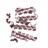
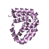
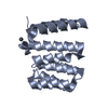
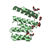
| Title | Fatty Acid and Retinol binding protein Na-FAR-1 from Necator americanus | ||||||||||||
 Components Components | Nematode fatty acid retinoid binding protein | ||||||||||||
 Keywords Keywords |  retinol-binding protein / Fatty acid retinol binding retinol-binding protein / Fatty acid retinol binding | ||||||||||||
| Function / homology | Nematode fatty acid retinoid binding / Nematode fatty acid retinoid binding protein (Gp-FAR-1) / lipid binding / extracellular region / PALMITIC ACID / Nematode fatty acid retinoid binding protein Function and homology information Function and homology information | ||||||||||||
| Biological species |  Necator americanus (New World hookworm) Necator americanus (New World hookworm) | ||||||||||||
| Method | X-RAY DIFFRACTION / SYNCHROTRON / SAD / Resolution: 2.14 Å | ||||||||||||
 Authors Authors | Gabrielsen, M. / Rey-Burusco, M.F. / Ibanez-Shimabukuro, M. / Griffiths, K. / Kennedy, M.W. / Corsico, B. / Smith, B.O. | ||||||||||||
| Funding support |  United Kingdom, United Kingdom,  Argentina, 3items Argentina, 3items
| ||||||||||||
 Citation Citation | Journal: Biochem.J. / Year: 2015 Title: Diversity in the structures and ligand-binding sites of nematode fatty acid and retinol-binding proteins revealed by Na-FAR-1 from Necator americanus. Authors: Rey-Burusco, M.F. / Ibanez-Shimabukuro, M. / Gabrielsen, M. / Franchini, G.R. / Roe, A.J. / Griffiths, K. / Zhan, B. / Cooper, A. / Kennedy, M.W. / Corsico, B. / Smith, B.O. | ||||||||||||
| History |
|
- Structure visualization
| Structure viewer | Molecule: MolmilJmol/JSmol |
|---|
- Downloads & links
-Download
| PDBx/mmCIF format | 4xcp.cif.gz | 75.3 KB | Display | PDBx/mmCIF format |
|---|---|---|---|---|
| PDB format | pdb4xcp.ent.gz | 60.6 KB | Display | PDB format |
| PDBx/mmJSON format | 4xcp.json.gz | Tree view | PDBx/mmJSON format | |
| Others |  Other downloads Other downloads |
-Validation report
| Arichive directory | https://data.pdbj.org/pub/pdb/validation_reports/xc/4xcpftp://data.pdbj.org/pub/pdb/validation_reports/xc/4xcp | HTTPS FTP |
|---|
-Related structure data


-Links
 PDBj
PDBj


- Assembly
| Deposited unit | 
| ||||||||
|---|---|---|---|---|---|---|---|---|---|
| 1 |
| ||||||||
| Unit cell |
| ||||||||
| Components on special symmetry positions |
|
-Components
| #1: Protein | Mass: 19038.008 Da / Num. of mol.: 1 / Fragment: UNP residues 21-175 / Mutation: M1MSE, M15MSE, M70MSE, M115MSE Source method: isolated from a genetically manipulated source Source: (gene. exp.) Necator americanus (New World hookworm)Gene: NECAME_14208 / Plasmid: pET11a / Production host:  Escherichia coli (E. coli) / Strain (production host): B834 / References: UniProt: W2SRJ3 Escherichia coli (E. coli) / Strain (production host): B834 / References: UniProt: W2SRJ3 | ||
|---|---|---|---|
| #2: Chemical | Palmitic acid  Mass: 256.424 Da / Num. of mol.: 2 / Source method: obtained synthetically / Formula: C16H32O2 Mass: 256.424 Da / Num. of mol.: 2 / Source method: obtained synthetically / Formula: C16H32O2#3: Water | ChemComp-HOH / | Water Mass: 18.015 Da / Num. of mol.: 136 / Source method: isolated from a natural source / Formula: H2O Mass: 18.015 Da / Num. of mol.: 136 / Source method: isolated from a natural source / Formula: H2O |
-Experimental details
-Experiment
| Experiment | Method: X-RAY DIFFRACTION |
|---|
- Sample preparation
| Crystal | Density Matthews: 4.48 Å3/Da / Density % sol: 72.53 % / Description: Cubic |
|---|---|
| Crystal grow | Temperature: 293 K / Method: vapor diffusion, sitting drop / pH: 4.2 / Details: 38 % PEG 300, 100 mM phosphate citrate pH 4.2 |
-Data collection
| Diffraction | Mean temperature: 100 K |
|---|---|
| Diffraction source | Source: SYNCHROTRON / Site: Diamond / Beamline: I04 / Wavelength: 0.9793 Å |
| Detector | Type: DECTRIS PILATUS 6M-F / Detector: PIXEL / Date: Dec 14, 2011 |
| Radiation | Protocol: SINGLE WAVELENGTH / Monochromatic (M) / Laue (L): M / Scattering type: x-ray |
| Radiation wavelength | Wavelength: 0.9793 Å / Relative weight: 1 |
| Reflection | Resolution: 2.14→29.38 Å / Num. all: 17353 / Num. obs: 17353 / % possible obs: 99.91 % / Redundancy: 28.9 % / Biso Wilson estimate: 30.11 Å2 / Rmerge(I) obs: 0.142 / Net I/σ(I): 25.5 |
| Reflection shell | Resolution: 2.14→2.27 Å / Redundancy: 14.5 % / Rmerge(I) obs: 0.785 / Mean I/σ(I) obs: 4.2 / % possible all: 97 |
- Processing
| Software |
| ||||||||||||||||||||||||||||||||||||||||||||||||||||||||||||||||||||||||||||||||||||||||||||||||||||||||||||||||||
|---|---|---|---|---|---|---|---|---|---|---|---|---|---|---|---|---|---|---|---|---|---|---|---|---|---|---|---|---|---|---|---|---|---|---|---|---|---|---|---|---|---|---|---|---|---|---|---|---|---|---|---|---|---|---|---|---|---|---|---|---|---|---|---|---|---|---|---|---|---|---|---|---|---|---|---|---|---|---|---|---|---|---|---|---|---|---|---|---|---|---|---|---|---|---|---|---|---|---|---|---|---|---|---|---|---|---|---|---|---|---|---|---|---|---|---|
| Refinement | Method to determine structure: SAD / Resolution: 2.14→29.38 Å / Cor.coef. Fo:Fc: 0.9252 / Cor.coef. Fo:Fc free: 0.9201 / SU R Cruickshank DPI: 0.149 / Cross valid method: THROUGHOUT / σ(F): 0 / SU R Blow DPI: 0.16 / SU Rfree Blow DPI: 0.14 / SU Rfree Cruickshank DPI: 0.134
| ||||||||||||||||||||||||||||||||||||||||||||||||||||||||||||||||||||||||||||||||||||||||||||||||||||||||||||||||||
| Displacement parameters | Biso mean: 32.54 Å2
| ||||||||||||||||||||||||||||||||||||||||||||||||||||||||||||||||||||||||||||||||||||||||||||||||||||||||||||||||||
| Refine analyze | Luzzati coordinate error obs: 0.273 Å | ||||||||||||||||||||||||||||||||||||||||||||||||||||||||||||||||||||||||||||||||||||||||||||||||||||||||||||||||||
| Refinement step | Cycle: LAST / Resolution: 2.14→29.38 Å
| ||||||||||||||||||||||||||||||||||||||||||||||||||||||||||||||||||||||||||||||||||||||||||||||||||||||||||||||||||
| Refine LS restraints |
| ||||||||||||||||||||||||||||||||||||||||||||||||||||||||||||||||||||||||||||||||||||||||||||||||||||||||||||||||||
| LS refinement shell | Resolution: 2.14→2.27 Å / Total num. of bins used: 9
| ||||||||||||||||||||||||||||||||||||||||||||||||||||||||||||||||||||||||||||||||||||||||||||||||||||||||||||||||||
| Refinement TLS params. | Method: refined / Origin x: 39.7537 Å / Origin y: 30.7686 Å / Origin z: 11.0724 Å
| ||||||||||||||||||||||||||||||||||||||||||||||||||||||||||||||||||||||||||||||||||||||||||||||||||||||||||||||||||
| Refinement TLS group | Selection details: { A|* } |
