coagulation factor VIIa / response to Thyroid stimulating hormone / response to 2,3,7,8-tetrachlorodibenzodioxine / response to astaxanthin / response to thyrotropin-releasing hormone / response to genistein / serine-type peptidase complex / positive regulation of platelet-derived growth factor receptor signaling pathway / response to vitamin K / response to carbon dioxide ...coagulation factor VIIa / response to Thyroid stimulating hormone / response to 2,3,7,8-tetrachlorodibenzodioxine / response to astaxanthin / response to thyrotropin-releasing hormone / response to genistein / serine-type peptidase complex / positive regulation of platelet-derived growth factor receptor signaling pathway / response to vitamin K / response to carbon dioxide / response to thyroxine / positive regulation of leukocyte chemotaxis / response to cholesterol / response to growth hormone / positive regulation of positive chemotaxis / Extrinsic Pathway of Fibrin Clot Formation / positive regulation of TOR signaling / positive regulation of blood coagulation / animal organ regeneration / Gamma-carboxylation of protein precursors / Transport of gamma-carboxylated protein precursors from the endoplasmic reticulum to the Golgi apparatus / Removal of aminoterminal propeptides from gamma-carboxylated proteins / serine-type peptidase activity / BMAL1:CLOCK,NPAS2 activates circadian gene expression / protein processing / Golgi lumen / circadian rhythm / response to estrogen / blood coagulation / response to estradiol / collagen-containing extracellular matrix / vesicle / response to hypoxia / positive regulation of cell migration / endoplasmic reticulum lumen / signaling receptor binding / serine-type endopeptidase activity / calcium ion binding / extracellular space / extracellular region / plasma membrane Similarity search - Function
In the structure databanks used in Yorodumi, some data are registered as the other names, "COVID-19 virus" and "2019-nCoV". Here are the details of the virus and the list of structure data.
Jan 31, 2019. EMDB accession codes are about to change! (news from PDBe EMDB page)
EMDB accession codes are about to change! (news from PDBe EMDB page)
The allocation of 4 digits for EMDB accession codes will soon come to an end. Whilst these codes will remain in use, new EMDB accession codes will include an additional digit and will expand incrementally as the available range of codes is exhausted. The current 4-digit format prefixed with “EMD-” (i.e. EMD-XXXX) will advance to a 5-digit format (i.e. EMD-XXXXX), and so on. It is currently estimated that the 4-digit codes will be depleted around Spring 2019, at which point the 5-digit format will come into force.
The EM Navigator/Yorodumi systems omit the EMD- prefix.
Related info.:Q: What is EMD? / ID/Accession-code notation in Yorodumi/EM Navigator
Yorodumi is a browser for structure data from EMDB, PDB, SASBDB, etc.
This page is also the successor to EM Navigator detail page, and also detail information page/front-end page for Omokage search.
The word "yorodu" (or yorozu) is an old Japanese word meaning "ten thousand". "mi" (miru) is to see.
Related info.:EMDB / PDB / SASBDB / Comparison of 3 databanks / Yorodumi Search / Aug 31, 2016. New EM Navigator & Yorodumi / Yorodumi Papers / Jmol/JSmol / Function and homology information / Changes in new EM Navigator and Yorodumi
 Movie
Movie Controller

 Yorodumi
 Open data
 Basic information
 Components
 Keywords
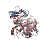
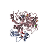
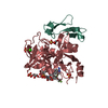
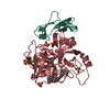
 GLYCOPROTEIN /
  Function and homology information
 Authors
 Citation
 Structure visualization
 Downloads & links
 Other downloads


 PDBj
PDBj


 Assembly
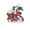

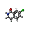


 Mass: 181.619 Da / Num. of mol.: 1 / Source method: obtained synthetically / Formula: C9H8ClNO
 Mass: 40.078 Da / Num. of mol.: 1 / Source method: obtained synthetically / Formula: Ca
 Mass: 96.063 Da / Num. of mol.: 3 / Source method: obtained synthetically / Formula: SO4
 Mass: 92.094 Da / Num. of mol.: 1 / Source method: obtained synthetically / Formula: C3H8O3
 Sample preparation
 / Beamline: 17-ID / Wavelength: 1 Å
 Processing