 Movie
Movie Controller

[English] 日本語
 Yorodumi
- PDB-4x6i: Development of N-(Functionalized benzoyl)-homocycloleucyl-glycino... -
+ Open data

- Basic information
| Entry | Database: PDB / ID: 4x6i | ||||||
|---|---|---|---|---|---|---|---|
| Title | Development of N-(Functionalized benzoyl)-homocycloleucyl-glycinonitriles as Potent Cathepsin K Inhibitors. | ||||||
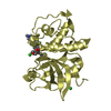
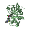
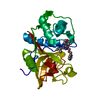
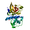
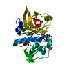
 Components Components | Cathepsin K | ||||||
 Keywords Keywords | HYDROLASE / cathepsin K / inhibitor | ||||||
| Function / homology |  Function and homology informationcathepsin K / mononuclear cell differentiation / intramembranous ossification / negative regulation of cartilage development / cellular response to zinc ion starvation / RUNX1 regulates transcription of genes involved in differentiation of keratinocytes / thyroid hormone generation / endolysosome lumen / Trafficking and processing of endosomal TLR / proteoglycan binding ...cathepsin K / mononuclear cell differentiation / intramembranous ossification / negative regulation of cartilage development / cellular response to zinc ion starvation / RUNX1 regulates transcription of genes involved in differentiation of keratinocytes / thyroid hormone generation / endolysosome lumen / Trafficking and processing of endosomal TLR / proteoglycan binding / Activation of Matrix Metalloproteinases / cysteine-type endopeptidase activator activity involved in apoptotic process / mitophagy / Collagen degradation / fibronectin binding / collagen catabolic process / extracellular matrix disassembly / cysteine-type peptidase activity / positive regulation of apoptotic signaling pathway / bone resorption / cellular response to transforming growth factor beta stimulus / collagen binding / MHC class II antigen presentation / Degradation of the extracellular matrix / proteolysis involved in protein catabolic process / lysosomal lumen / response to insulin / response to organic cyclic compound / cellular response to tumor necrosis factor / response to ethanol / lysosome / immune response / apical plasma membrane / external side of plasma membrane / cysteine-type endopeptidase activity / serine-type endopeptidase activity / intracellular membrane-bounded organelle / proteolysis / extracellular space / extracellular region / nucleoplasm Function and homology informationcathepsin K / mononuclear cell differentiation / intramembranous ossification / negative regulation of cartilage development / cellular response to zinc ion starvation / RUNX1 regulates transcription of genes involved in differentiation of keratinocytes / thyroid hormone generation / endolysosome lumen / Trafficking and processing of endosomal TLR / proteoglycan binding ...cathepsin K / mononuclear cell differentiation / intramembranous ossification / negative regulation of cartilage development / cellular response to zinc ion starvation / RUNX1 regulates transcription of genes involved in differentiation of keratinocytes / thyroid hormone generation / endolysosome lumen / Trafficking and processing of endosomal TLR / proteoglycan binding / Activation of Matrix Metalloproteinases / cysteine-type endopeptidase activator activity involved in apoptotic process / mitophagy / Collagen degradation / fibronectin binding / collagen catabolic process / extracellular matrix disassembly / cysteine-type peptidase activity / positive regulation of apoptotic signaling pathway / bone resorption / cellular response to transforming growth factor beta stimulus / collagen binding / MHC class II antigen presentation / Degradation of the extracellular matrix / proteolysis involved in protein catabolic process / lysosomal lumen / response to insulin / response to organic cyclic compound / cellular response to tumor necrosis factor / response to ethanol / lysosome / immune response / apical plasma membrane / external side of plasma membrane / cysteine-type endopeptidase activity / serine-type endopeptidase activity / intracellular membrane-bounded organelle / proteolysis / extracellular space / extracellular region / nucleoplasmSimilarity search - Function | ||||||
| Biological species |  Homo sapiens (human) Homo sapiens (human) | ||||||
| Method | X-RAY DIFFRACTION / MOLECULAR REPLACEMENT / molecular replacement / Resolution: 1.87 Å | ||||||
 Authors Authors | Borisek, J. / Mohar, B. / Vizovisek, M. / Sosnowski, P. / Turk, D. / Turk, B. / Novic, M. | ||||||
 Citation Citation | Journal: J.Med.Chem. / Year: 2015 Title: Development of N-(Functionalized benzoyl)-homocycloleucyl-glycinonitriles as Potent Cathepsin K Inhibitors. Authors: Borisek, J. / Vizovisek, M. / Sosnowski, P. / Turk, B. / Turk, D. / Mohar, B. / Novic, M. | ||||||
| History |
|
- Structure visualization
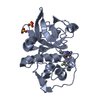
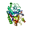
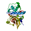
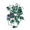
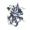
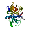
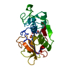
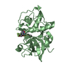
| Structure viewer | Molecule: MolmilJmol/JSmol |
|---|
- Downloads & links
-Download
| PDBx/mmCIF format | 4x6i.cif.gz | 61.3 KB | Display | PDBx/mmCIF format |
|---|---|---|---|---|
| PDB format | pdb4x6i.ent.gz | 43 KB | Display | PDB format |
| PDBx/mmJSON format | 4x6i.json.gz | Tree view | PDBx/mmJSON format | |
| Others |  Other downloads Other downloads |
-Validation report
| Arichive directory | https://data.pdbj.org/pub/pdb/validation_reports/x6/4x6iftp://data.pdbj.org/pub/pdb/validation_reports/x6/4x6i | HTTPS FTP |
|---|
-Related structure data
| Related structure data | 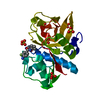 4x6hC  4x6jC 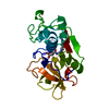 1atkS S: Starting model for refinement C: citing same article ( |
|---|---|
| Similar structure data |


-Links
 PDBj
PDBj


- Assembly
| Deposited unit | 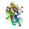
| ||||||||
|---|---|---|---|---|---|---|---|---|---|
| 1 |
| ||||||||
| Unit cell |
|
-Components
| #1: Protein | / Cathepsin O / Cathepsin O2 / Cathepsin X Mass: 23523.480 Da / Num. of mol.: 1 / Fragment: UNP RESIDUES 115-329 Source method: isolated from a genetically manipulated source Source: (gene. exp.) Homo sapiens (human) / Gene: CTSK, CTSO, CTSO2 / Production host:  Komagataella pastoris GS115 (fungus) / References: UniProt: P43235, cathepsin K Komagataella pastoris GS115 (fungus) / References: UniProt: P43235, cathepsin K | ||
|---|---|---|---|
| #2: Chemical | ChemComp-3Y1 / 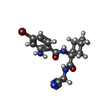  Mass: 379.252 Da / Num. of mol.: 1 / Source method: obtained synthetically / Formula: C16H19BrN4O2 Mass: 379.252 Da / Num. of mol.: 1 / Source method: obtained synthetically / Formula: C16H19BrN4O2 | ||
| #3: Chemical | ChemComp-SO4 / Sulfate  Mass: 96.063 Da / Num. of mol.: 5 / Source method: obtained synthetically / Formula: SO4 Mass: 96.063 Da / Num. of mol.: 5 / Source method: obtained synthetically / Formula: SO4#4: Water | ChemComp-HOH / | Water Mass: 18.015 Da / Num. of mol.: 201 / Source method: isolated from a natural source / Formula: H2O Mass: 18.015 Da / Num. of mol.: 201 / Source method: isolated from a natural source / Formula: H2O |
-Experimental details
-Experiment
| Experiment | Method: X-RAY DIFFRACTION / Number of used crystals: 1 |
|---|
- Sample preparation
| Crystal | Density Matthews: 1.85 Å3/Da / Density % sol: 33.65 % |
|---|---|
| Crystal grow | Temperature: 295 K / Method: vapor diffusion, sitting drop Details: Lithium sulfate 0.2M, Sodium acetate 0.1M, Peg 8000 30% |
-Data collection
| Diffraction | Mean temperature: 100 K |
|---|---|
| Diffraction source | Source: ROTATING ANODE / Type: BRUKER AXS MICROSTAR / Wavelength: 1.54184 Å |
| Detector | Type: Bruker Platinum 135 / Detector: CCD / Date: Jan 5, 2014 |
| Radiation | Protocol: SINGLE WAVELENGTH / Monochromatic (M) / Laue (L): M / Scattering type: x-ray |
| Radiation wavelength | Wavelength: 1.54184 Å / Relative weight: 1 |
| Reflection | Resolution: 1.87→46.28 Å / Num. obs: 14804 / % possible obs: 98.31 % / Redundancy: 9.5 % / Rmerge(I) obs: 0.2593 / Net I/σ(I): 9.96 |
| Reflection shell | Resolution: 1.87→1.937 Å / Redundancy: 7.4 % / Rmerge(I) obs: 0.9815 / % possible all: 98.16 |
-Phasing
| Phasing | Method: molecular replacement | |||||||||
|---|---|---|---|---|---|---|---|---|---|---|
| Phasing MR | Model details: Phaser MODE: MR_AUTO
|
- Processing
| Software |
| |||||||||||||||||||||||||||||||||||||||||||||||||||||||||||||||||||||||||||
|---|---|---|---|---|---|---|---|---|---|---|---|---|---|---|---|---|---|---|---|---|---|---|---|---|---|---|---|---|---|---|---|---|---|---|---|---|---|---|---|---|---|---|---|---|---|---|---|---|---|---|---|---|---|---|---|---|---|---|---|---|---|---|---|---|---|---|---|---|---|---|---|---|---|---|---|---|
| Refinement | Method to determine structure: MOLECULAR REPLACEMENT Starting model: 1ATK Resolution: 1.87→46.28 Å / Cor.coef. Fo:Fc: 0.931 / Cor.coef. Fo:Fc free: 0.888 / WRfactor Rfree: 0.1988 / WRfactor Rwork: 0.1549 / FOM work R set: 0.8063 / SU B: 4.162 / SU ML: 0.12 / SU R Cruickshank DPI: 0.1887 / SU Rfree: 0.1671 / Cross valid method: THROUGHOUT / σ(F): 0 / ESU R: 0.189 / ESU R Free: 0.167 / Stereochemistry target values: MAXIMUM LIKELIHOOD Details: HYDROGENS HAVE BEEN ADDED IN THE RIDING POSITIONS U VALUES : REFINED INDIVIDUALLY
| |||||||||||||||||||||||||||||||||||||||||||||||||||||||||||||||||||||||||||
| Solvent computation | Ion probe radii: 0.8 Å / Shrinkage radii: 0.8 Å / VDW probe radii: 1.2 Å / Solvent model: MASK | |||||||||||||||||||||||||||||||||||||||||||||||||||||||||||||||||||||||||||
| Displacement parameters | Biso max: 36.95 Å2 / Biso mean: 8.629 Å2 / Biso min: 2 Å2
| |||||||||||||||||||||||||||||||||||||||||||||||||||||||||||||||||||||||||||
| Refinement step | Cycle: final / Resolution: 1.87→46.28 Å
| |||||||||||||||||||||||||||||||||||||||||||||||||||||||||||||||||||||||||||
| Refine LS restraints |
| |||||||||||||||||||||||||||||||||||||||||||||||||||||||||||||||||||||||||||
| LS refinement shell | Resolution: 1.87→1.919 Å / Total num. of bins used: 20
|
