 Movie
Movie Controller

[English] 日本語
 Yorodumi
- PDB-4w8d: Crystal structure of MST3 with a pyrrolopyrimidine inhibitor (PF-... -
+ Open data

- Basic information
| Entry | Database: PDB / ID: 4w8d | ||||||
|---|---|---|---|---|---|---|---|
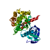
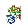
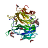
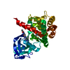
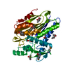
| Title | Crystal structure of MST3 with a pyrrolopyrimidine inhibitor (PF-06454589). | ||||||
 Components Components | Serine/threonine-protein kinase 24 36 kDa subunit | ||||||
 Keywords Keywords | transferase/transferase inhibitor / MST3 / Pyrrolopyrimidine /  Kinase / inhibitor / transferase-transferase inhibitor complex Kinase / inhibitor / transferase-transferase inhibitor complex | ||||||
| Function / homology |  Function and homology information Function and homology informationApoptotic execution phase / regulation of axon regeneration / execution phase of apoptosis / intrinsic apoptotic signaling pathway in response to oxidative stress / positive regulation of axon regeneration / Apoptotic cleavage of cellular proteins / cellular response to starvation / negative regulation of cell migration / response to hydrogen peroxide / protein autophosphorylation ...Apoptotic execution phase / regulation of axon regeneration / execution phase of apoptosis / intrinsic apoptotic signaling pathway in response to oxidative stress / positive regulation of axon regeneration / Apoptotic cleavage of cellular proteins / cellular response to starvation / negative regulation of cell migration / response to hydrogen peroxide / protein autophosphorylation / non-specific serine/threonine protein kinase / protein kinase activity / cadherin binding / Golgi membrane / protein phosphorylation / protein serine kinase activity / protein serine/threonine kinase activity / nucleolus / Golgi apparatus / signal transduction / extracellular exosome / nucleoplasm / ATP binding / metal ion binding / nucleus / cytosol / cytoplasmSimilarity search - Function | ||||||
| Biological species |  Homo sapiens (human) Homo sapiens (human) | ||||||
| Method | X-RAY DIFFRACTION / SYNCHROTRON / Resolution: 1.77 Å | ||||||
 Authors Authors | Jasti, J. / Song, X. / Griffor, M. / Kurumbail, R.G. | ||||||
 Citation Citation | Journal: J.Med.Chem. / Year: 2015 Title: Discovery and preclinical profiling of 3-[4-(morpholin-4-yl)-7H-pyrrolo[2,3-d]pyrimidin-5-yl]benzonitrile (PF-06447475), a highly potent, selective, brain penetrant, and in vivo active LRRK2 kinase inhibitor. Authors: Henderson, J.L. / Kormos, B.L. / Hayward, M.M. / Coffman, K.J. / Jasti, J. / Kurumbail, R.G. / Wager, T.T. / Verhoest, P.R. / Noell, G.S. / Chen, Y. / Needle, E. / Berger, Z. / Steyn, S.J. / ...Authors: Henderson, J.L. / Kormos, B.L. / Hayward, M.M. / Coffman, K.J. / Jasti, J. / Kurumbail, R.G. / Wager, T.T. / Verhoest, P.R. / Noell, G.S. / Chen, Y. / Needle, E. / Berger, Z. / Steyn, S.J. / Houle, C. / Hirst, W.D. / Galatsis, P. | ||||||
| History |
|
- Structure visualization
| Structure viewer | Molecule: MolmilJmol/JSmol |
|---|
- Downloads & links
-Download
| PDBx/mmCIF format | 4w8d.cif.gz | 80.2 KB | Display | PDBx/mmCIF format |
|---|---|---|---|---|
| PDB format | pdb4w8d.ent.gz | 58.5 KB | Display | PDB format |
| PDBx/mmJSON format | 4w8d.json.gz | Tree view | PDBx/mmJSON format | |
| Others |  Other downloads Other downloads |
-Validation report
| Arichive directory | https://data.pdbj.org/pub/pdb/validation_reports/w8/4w8dftp://data.pdbj.org/pub/pdb/validation_reports/w8/4w8d | HTTPS FTP |
|---|
-Related structure data


-Links
 PDBj
PDBj
- Assembly
| Deposited unit | 
| ||||||||
|---|---|---|---|---|---|---|---|---|---|
| 1 |
| ||||||||
| Unit cell |
| ||||||||
| Components on special symmetry positions |
|
-Components
| #1: Protein | Mass: 32954.770 Da / Num. of mol.: 1 / Fragment: UNP residues 9-298 Source method: isolated from a genetically manipulated source Source: (gene. exp.) Homo sapiens (human) / Gene: STK24, MST3, STK3 / Production host:  Escherichia coli (E. coli) Escherichia coli (E. coli)References: UniProt: Q9Y6E0, non-specific serine/threonine protein kinase |
|---|---|
| #2: Chemical | ChemComp-3JA /   Mass: 284.316 Da / Num. of mol.: 1 / Source method: obtained synthetically / Formula: C14H16N6O Mass: 284.316 Da / Num. of mol.: 1 / Source method: obtained synthetically / Formula: C14H16N6O |
| #3: Water | ChemComp-HOH / Water Mass: 18.015 Da / Num. of mol.: 291 / Source method: isolated from a natural source / Formula: H2O Mass: 18.015 Da / Num. of mol.: 291 / Source method: isolated from a natural source / Formula: H2O |
-Experimental details
-Experiment
| Experiment | Method: X-RAY DIFFRACTION / Number of used crystals: 1 |
|---|
- Sample preparation
| Crystal | Density Matthews: 2.74 Å3/Da / Density % sol: 55.15 % |
|---|---|
| Crystal grow | Temperature: 293 K / Method: vapor diffusion, sitting drop / pH: 8.5 Details: 0.1M Tris-HCL (pH 8.5), 180mM MgCl2, 5mM Manganese acetate, 10-17% PEG 3350 |
-Data collection
| Diffraction | Mean temperature: 100 K |
|---|---|
| Diffraction source | Source: SYNCHROTRON / Site: APS  / Beamline: 17-ID / Wavelength: 1 Å / Beamline: 17-ID / Wavelength: 1 Å |
| Detector | Type: DECTRIS PILATUS 6M / Detector: PIXEL / Date: Jun 27, 2012 |
| Radiation | Protocol: SINGLE WAVELENGTH / Monochromatic (M) / Laue (L): M / Scattering type: x-ray |
| Radiation wavelength | Wavelength: 1 Å / Relative weight: 1 |
| Reflection | Resolution: 1.77→49.88 Å / Num. obs: 34094 / % possible obs: 97.1 % / Redundancy: 3 % / Biso Wilson estimate: 31.55 Å2 / Rsym value: 0.034 / Net I/σ(I): 16.2 |
- Processing
| Software | Name: BUSTER / Version: 2.11.2 / Classification: refinement | ||||||||||||||||||||||||||||||||||||||||||||||||||||||||||||||||||||||||||||||||||||||||||||||||||||||||||||||||||
|---|---|---|---|---|---|---|---|---|---|---|---|---|---|---|---|---|---|---|---|---|---|---|---|---|---|---|---|---|---|---|---|---|---|---|---|---|---|---|---|---|---|---|---|---|---|---|---|---|---|---|---|---|---|---|---|---|---|---|---|---|---|---|---|---|---|---|---|---|---|---|---|---|---|---|---|---|---|---|---|---|---|---|---|---|---|---|---|---|---|---|---|---|---|---|---|---|---|---|---|---|---|---|---|---|---|---|---|---|---|---|---|---|---|---|---|
| Refinement | Resolution: 1.77→40.16 Å / Cor.coef. Fo:Fc: 0.9586 / Cor.coef. Fo:Fc free: 0.9586 / SU R Cruickshank DPI: 0.102 / Cross valid method: THROUGHOUT / σ(F): 0 / SU R Blow DPI: 0.106 / SU Rfree Blow DPI: 0.093 / SU Rfree Cruickshank DPI: 0.091
| ||||||||||||||||||||||||||||||||||||||||||||||||||||||||||||||||||||||||||||||||||||||||||||||||||||||||||||||||||
| Displacement parameters | Biso mean: 37.84 Å2
| ||||||||||||||||||||||||||||||||||||||||||||||||||||||||||||||||||||||||||||||||||||||||||||||||||||||||||||||||||
| Refine analyze | Luzzati coordinate error obs: 0.191 Å | ||||||||||||||||||||||||||||||||||||||||||||||||||||||||||||||||||||||||||||||||||||||||||||||||||||||||||||||||||
| Refinement step | Cycle: 1 / Resolution: 1.77→40.16 Å
| ||||||||||||||||||||||||||||||||||||||||||||||||||||||||||||||||||||||||||||||||||||||||||||||||||||||||||||||||||
| Refine LS restraints |
| ||||||||||||||||||||||||||||||||||||||||||||||||||||||||||||||||||||||||||||||||||||||||||||||||||||||||||||||||||
| LS refinement shell | Resolution: 1.77→1.82 Å / Total num. of bins used: 17
|
