 Movie
Movie Controller


+ Open data

- Basic information
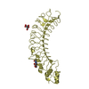
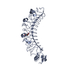
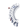
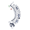
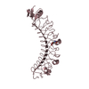
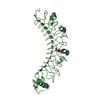
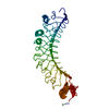
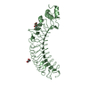
| Entry | Database: PDB / ID: 4v2e | ||||||
|---|---|---|---|---|---|---|---|
| Title | FLRT3 LRR domain | ||||||
 Components Components | FIBRONECTIN LEUCINE RICH TRANSMEMBRANE PROTEIN 3 | ||||||
 Keywords Keywords |  SIGNALING PROTEIN / LEUCINE-RICH REPEAT / LRR / UNC5 SIGNALING PROTEIN / LEUCINE-RICH REPEAT / LRR / UNC5 | ||||||
| Function / homology |  Function and homology information Function and homology informationproepicardium cell migration involved in pericardium morphogenesis / Downstream signaling of activated FGFR1 / head development / synaptic membrane adhesion / growth cone membrane / cell-cell adhesion via plasma-membrane adhesion molecules / embryonic morphogenesis / fibroblast growth factor receptor binding / chemorepellent activity / positive regulation of synapse assembly ...proepicardium cell migration involved in pericardium morphogenesis / Downstream signaling of activated FGFR1 / head development / synaptic membrane adhesion / growth cone membrane / cell-cell adhesion via plasma-membrane adhesion molecules / embryonic morphogenesis / fibroblast growth factor receptor binding / chemorepellent activity / positive regulation of synapse assembly / neuron projection extension / response to axon injury / fibroblast growth factor receptor signaling pathway / axon terminus / axonal growth cone / synapse assembly / synaptic membrane / axon guidance / synapse organization / neuron projection development / cell-cell junction / cell junction / heart development / postsynaptic membrane / postsynaptic density / focal adhesion / glutamatergic synapse / endoplasmic reticulum membrane / protein homodimerization activity / extracellular space / plasma membrane / cytosolSimilarity search - Function | ||||||
| Biological species |  MUS MUSCULUS (house mouse) MUS MUSCULUS (house mouse) | ||||||
| Method | X-RAY DIFFRACTION / SYNCHROTRON / MOLECULAR REPLACEMENT / Resolution: 2.5 Å | ||||||
 Authors Authors | Seiradake, E. / del Toro, D. / Nagel, D. / Cop, F. / Haertl, R. / Ruff, T. / Seyit-Bremer, G. / Harlos, K. / Border, E.C. / Acker-Palmer, A. ...Seiradake, E. / del Toro, D. / Nagel, D. / Cop, F. / Haertl, R. / Ruff, T. / Seyit-Bremer, G. / Harlos, K. / Border, E.C. / Acker-Palmer, A. / Jones, E.Y. / Klein, R. | ||||||
 Citation Citation | Journal: Neuron / Year: 2014 Title: Flrt Structure: Balancing Repulsion and Cell Adhesion in Cortical and Vascular Development. Authors: Seiradake, E. / Del Toro, D. / Nagel, D. / Cop, F. / Hartl, R. / Ruff, T. / Seyit-Bremer, G. / Harlos, K. / Border, E.C. / Acker-Palmer, A. / Jones, E.Y. / Klein, R. | ||||||
| History |
|
- Structure visualization
| Structure viewer | Molecule: MolmilJmol/JSmol |
|---|
- Downloads & links
-Download
| PDBx/mmCIF format | 4v2e.cif.gz | 258.4 KB | Display | PDBx/mmCIF format |
|---|---|---|---|---|
| PDB format | pdb4v2e.ent.gz | 212 KB | Display | PDB format |
| PDBx/mmJSON format | 4v2e.json.gz | Tree view | PDBx/mmJSON format | |
| Others |  Other downloads Other downloads |
-Validation report
| Arichive directory | https://data.pdbj.org/pub/pdb/validation_reports/v2/4v2eftp://data.pdbj.org/pub/pdb/validation_reports/v2/4v2e | HTTPS FTP |
|---|
-Related structure data
| Related structure data | 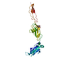 4v2aC 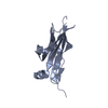 4v2bC  4v2cC  4v2dSC C: citing same article ( S: Starting model for refinement |
|---|---|
| Similar structure data |


-Links
 PDBj
PDBj


- Assembly
| Deposited unit | 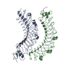
| ||||||||
|---|---|---|---|---|---|---|---|---|---|
| 1 | 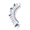
| ||||||||
| 2 | 
| ||||||||
| Unit cell |
|
-Components
| #1: Protein | Mass: 37557.832 Da / Num. of mol.: 2 / Fragment: LRR DOMAIN Source method: isolated from a genetically manipulated source Source: (gene. exp.) MUS MUSCULUS (house mouse) / Plasmid: PHLSEC / Cell line (production host): HEK293T / Production host:  HOMO SAPIENS (human) / References: UniProt: Q8BGT1 HOMO SAPIENS (human) / References: UniProt: Q8BGT1Sequence details | CONTAINS C-TERMINAL HIS TAG | |
|---|
-Experimental details
-Experiment
| Experiment | Method: X-RAY DIFFRACTION |
|---|
- Sample preparation
| Crystal | Density Matthews: 2.2 Å3/Da / Density % sol: 44.11 % / Description: NONE |
|---|
-Data collection
| Diffraction | Mean temperature: 100 K |
|---|---|
| Diffraction source | Source: SYNCHROTRON / Site: Diamond  / Beamline: I04 / Wavelength: 0.9611 / Beamline: I04 / Wavelength: 0.9611 |
| Detector | Type: DECTRIS PIXEL / Detector: PIXEL |
| Radiation | Protocol: SINGLE WAVELENGTH / Monochromatic (M) / Laue (L): M / Scattering type: x-ray |
| Radiation wavelength | Wavelength: 0.9611 Å / Relative weight: 1 |
| Reflection | Resolution: 2.5→57 Å / Num. obs: 63975 / % possible obs: 90.4 % / Redundancy: 3.1 % / Biso Wilson estimate: 35.05 Å2 / Rmerge(I) obs: 0.46 / Net I/σ(I): 3.1 |
| Reflection shell | Resolution: 2.5→2.6 Å / Redundancy: 2.7 % / Mean I/σ(I) obs: 0.98 / % possible all: 89.2 |
- Processing
| Software |
| ||||||||||||||||||||||||||||||||||||||||||||||||||||||||||||||||||||||||||||||||||||||||||||||||||||||||||||||||||
|---|---|---|---|---|---|---|---|---|---|---|---|---|---|---|---|---|---|---|---|---|---|---|---|---|---|---|---|---|---|---|---|---|---|---|---|---|---|---|---|---|---|---|---|---|---|---|---|---|---|---|---|---|---|---|---|---|---|---|---|---|---|---|---|---|---|---|---|---|---|---|---|---|---|---|---|---|---|---|---|---|---|---|---|---|---|---|---|---|---|---|---|---|---|---|---|---|---|---|---|---|---|---|---|---|---|---|---|---|---|---|---|---|---|---|---|
| Refinement | Method to determine structure: MOLECULAR REPLACEMENT Starting model: PDB ENTRY 4V2D Resolution: 2.5→57.38 Å / Cor.coef. Fo:Fc: 0.6342 / Cor.coef. Fo:Fc free: 0.6285 / SU R Cruickshank DPI: 5.742 / Cross valid method: THROUGHOUT / σ(F): 0 / SU R Blow DPI: 2.005 / SU Rfree Blow DPI: 0.435 / SU Rfree Cruickshank DPI: 0.452
| ||||||||||||||||||||||||||||||||||||||||||||||||||||||||||||||||||||||||||||||||||||||||||||||||||||||||||||||||||
| Displacement parameters | Biso mean: 40.66 Å2
| ||||||||||||||||||||||||||||||||||||||||||||||||||||||||||||||||||||||||||||||||||||||||||||||||||||||||||||||||||
| Refine analyze | Luzzati coordinate error obs: 1.212 Å | ||||||||||||||||||||||||||||||||||||||||||||||||||||||||||||||||||||||||||||||||||||||||||||||||||||||||||||||||||
| Refinement step | Cycle: LAST / Resolution: 2.5→57.38 Å
| ||||||||||||||||||||||||||||||||||||||||||||||||||||||||||||||||||||||||||||||||||||||||||||||||||||||||||||||||||
| Refine LS restraints |
| ||||||||||||||||||||||||||||||||||||||||||||||||||||||||||||||||||||||||||||||||||||||||||||||||||||||||||||||||||
| LS refinement shell | Resolution: 2.5→2.63 Å / Total num. of bins used: 10
| ||||||||||||||||||||||||||||||||||||||||||||||||||||||||||||||||||||||||||||||||||||||||||||||||||||||||||||||||||
| Refinement TLS params. | Method: refined / Refine-ID: X-RAY DIFFRACTION
|
