 Movie
Movie Controller

[English] 日本語
 Yorodumi
- PDB-4uyo: Structure of delta7-DgkA in 7.9 MAG by serial femtosecond crystat... -
+ Open data

- Basic information
| Entry | Database: PDB / ID: 4uyo | ||||||
|---|---|---|---|---|---|---|---|
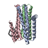
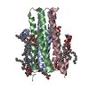
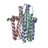
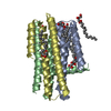
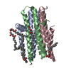
| Title | Structure of delta7-DgkA in 7.9 MAG by serial femtosecond crystatallography to 2.18 angstrom resolution | ||||||
 Components Components | DIACYLGLYCEROL KINASE-DELTA 7 | ||||||
 Keywords Keywords |  TRANSFERASE / 7.9 MAG / IN MESO CRYSTALLIZATION / LIPID CUBIC PHASE / LIPIDIC CUBIC PHASE / LIPID MESOPHASE / LIPIDIC MESOPHASE / MEMBRANE PROTEIN / MICROCRYSTALS / MONOACYLGLYCEROL / ROOM TEMPERATURE CRYSTALLOGRAPHY / SERIAL FEMTOSECOND CRYSTALLOGRAPHY / X-RAY FREE-ELECTRON LASER TRANSFERASE / 7.9 MAG / IN MESO CRYSTALLIZATION / LIPID CUBIC PHASE / LIPIDIC CUBIC PHASE / LIPID MESOPHASE / LIPIDIC MESOPHASE / MEMBRANE PROTEIN / MICROCRYSTALS / MONOACYLGLYCEROL / ROOM TEMPERATURE CRYSTALLOGRAPHY / SERIAL FEMTOSECOND CRYSTALLOGRAPHY / X-RAY FREE-ELECTRON LASER | ||||||
| Function / homology |  Function and homology information Function and homology informationdiacylglycerol kinase (ATP) / ATP-dependent diacylglycerol kinase activity / phosphatidic acid biosynthetic process / response to UV / phosphorylation / ATP binding / membrane / identical protein binding / metal ion binding / plasma membraneSimilarity search - Function | ||||||
| Biological species |  ESCHERICHIA COLI K12 (bacteria) ESCHERICHIA COLI K12 (bacteria) | ||||||
| Method | X-RAY DIFFRACTION / FREE ELECTRON LASER / MOLECULAR REPLACEMENT / Resolution: 2.18 Å | ||||||
 Authors Authors | Li, D. / Howe, N. / Other, O. / Caffrey, M. | ||||||
 Citation Citation | Journal: Nat.Commun. / Year: 2015 Title: Ternary Structure Reveals Mechanism of a Membrane Diacylglycerol Kinase. Authors: Li, D. / Stansfeld, P.J. / Sansom, M.S.P. / Keogh, A. / Vogeley, L. / Howe, N. / Lyons, J.A. / Aragao, D. / Fromme, P. / Fromme, R. / Basu, S. / Grotjohann, I. / Kupitz, C. / Rendek, K. / ...Authors: Li, D. / Stansfeld, P.J. / Sansom, M.S.P. / Keogh, A. / Vogeley, L. / Howe, N. / Lyons, J.A. / Aragao, D. / Fromme, P. / Fromme, R. / Basu, S. / Grotjohann, I. / Kupitz, C. / Rendek, K. / Weierstall, U. / Zatsepin, N.A. / Cherezov, V. / Liu, W. / Bandaru, S. / English, N.J. / Gati, C. / Barty, A. / Yefanov, O. / Chapman, H.N. / Diederichs, K. / Messerschmidt, M. / Boutet, S. / Williams, G.J. / Marvin Seibert, M. / Caffrey, M. | ||||||
| History |
|
- Structure visualization
| Structure viewer | Molecule: MolmilJmol/JSmol |
|---|
- Downloads & links
-Download
| PDBx/mmCIF format | 4uyo.cif.gz | 142.5 KB | Display | PDBx/mmCIF format |
|---|---|---|---|---|
| PDB format | pdb4uyo.ent.gz | 113.2 KB | Display | PDB format |
| PDBx/mmJSON format | 4uyo.json.gz | Tree view | PDBx/mmJSON format | |
| Others |  Other downloads Other downloads |
-Validation report
| Arichive directory | https://data.pdbj.org/pub/pdb/validation_reports/uy/4uyoftp://data.pdbj.org/pub/pdb/validation_reports/uy/4uyo | HTTPS FTP |
|---|
-Related structure data
| Related structure data |  4uxwC  4uxxC  4uxzC 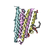 3ze3S C: citing same article ( S: Starting model for refinement |
|---|---|
| Similar structure data |


-Links
 PDBj
PDBj


- Assembly
| Deposited unit | 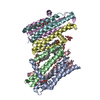
| ||||||||
|---|---|---|---|---|---|---|---|---|---|
| 1 | 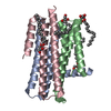
| ||||||||
| 2 | 
| ||||||||
| Unit cell |
|
-Components
-Protein , 1 types, 6 molecules ABCDEF
| #1: Protein | Mass: 14204.451 Da / Num. of mol.: 6 / Mutation: YES Source method: isolated from a genetically manipulated source Source: (gene. exp.) ESCHERICHIA COLI K12 (bacteria) / Plasmid: PTRCHISB-DGKA-DELTA7 / Production host: ESCHERICHIA COLI (E. coli) / Strain (production host): WH1061 / References: UniProt: P0ABN1, diacylglycerol kinase (ATP) |
|---|
-Non-polymers , 5 types, 96 molecules 


| #2: Chemical | ChemComp-79N / (  Mass: 328.487 Da / Num. of mol.: 5 / Source method: obtained synthetically / Formula: C19H36O4 Mass: 328.487 Da / Num. of mol.: 5 / Source method: obtained synthetically / Formula: C19H36O4#3: Chemical | ChemComp-79M / (  Mass: 328.487 Da / Num. of mol.: 4 / Source method: obtained synthetically / Formula: C19H36O4 Mass: 328.487 Da / Num. of mol.: 4 / Source method: obtained synthetically / Formula: C19H36O4#4: Chemical | ChemComp-ZN / |  Mass: 65.409 Da / Num. of mol.: 1 / Source method: obtained synthetically / Formula: Zn Mass: 65.409 Da / Num. of mol.: 1 / Source method: obtained synthetically / Formula: Zn#5: Chemical | ChemComp-FLC / | Citric acid Mass: 189.100 Da / Num. of mol.: 1 / Source method: obtained synthetically / Formula: C6H5O7 Mass: 189.100 Da / Num. of mol.: 1 / Source method: obtained synthetically / Formula: C6H5O7#6: Water | ChemComp-HOH / | WaterMass: 18.015 Da / Num. of mol.: 85 / Source method: isolated from a natural source / Formula: H2O |
|---|
-Experimental details
-Experiment
| Experiment | Method: X-RAY DIFFRACTION / Number of used crystals: 140440 |
|---|
- Sample preparation
| Crystal | Density Matthews: 2.93 Å3/Da / Density % sol: 58.04 % / Description: R SPLIT (I) 0.07, R SPLIT FOR SHELL (I) 2.38 |
|---|---|
| Crystal grow | Temperature: 293 K / Method: lipidic cubic phase / pH: 5.6 Details: 0.2 %(V/V) 2-METHYL-2, 4-PENTANEDIOL (MPD), 0.1 M SODIUM CHLORIDE, 0.1 M SODIUM CITRATE/HCL PH 5.6. CRYSTALLIZED USING THE IN MESO (LIPID CUBIC PHASE) METHOD AT 20 DEGREES CELCIUS WITH THE 7. ...Details: 0.2 %(V/V) 2-METHYL-2, 4-PENTANEDIOL (MPD), 0.1 M SODIUM CHLORIDE, 0.1 M SODIUM CITRATE/HCL PH 5.6. CRYSTALLIZED USING THE IN MESO (LIPID CUBIC PHASE) METHOD AT 20 DEGREES CELCIUS WITH THE 7.9 MONOACYLGLYCEROL (7.9 MAG) AS THE HOSTING LIPID.CRYSTALLIZATION WAS SET UP BY INCUBATING 20 MICROLITERS OF LIPID CUBIC PHASE WITH 400 MICROLITERS OF PRECIPITANT SOLUTION IN COUPLED SYRINGES. |
-Data collection
| Diffraction | Mean temperature: 293 K / Serial crystal experiment: Y |
|---|---|
| Diffraction source | Source: FREE ELECTRON LASER / Site: SLAC LCLS  / Beamline: CXI / Wavelength: 1.302 / Wavelength: 1.302 Å / Beamline: CXI / Wavelength: 1.302 / Wavelength: 1.302 Å |
| Detector | Type: Cornell-SLAC Pixel Array Detector (CSPAD) / Detector: PIXEL / Date: Mar 25, 2013 |
| Radiation | Monochromator: K-B MIRRORS / Protocol: SINGLE WAVELENGTH / Monochromatic (M) / Laue (L): M / Scattering type: x-ray |
| Radiation wavelength | Wavelength: 1.302 Å / Relative weight: 1 |
| Reflection | Resolution: 2.18→40.5 Å / Num. obs: 54058 / % possible obs: 100 % / Observed criterion σ(I): -5 / Redundancy: 3466 % / Biso Wilson estimate: 71.31 Å2 / Net I/σ(I): 19 |
| Reflection shell | Resolution: 2.18→2.24 Å / Redundancy: 3519 % / Mean I/σ(I) obs: 0.44 / % possible all: 100 |
- Processing
| Software |
| |||||||||||||||||||||||||||||||||||||||||||||||||||||||||||||||||||||||||||||||||||||||||||||||||||||||||||||||||||||||||||||||||||||
|---|---|---|---|---|---|---|---|---|---|---|---|---|---|---|---|---|---|---|---|---|---|---|---|---|---|---|---|---|---|---|---|---|---|---|---|---|---|---|---|---|---|---|---|---|---|---|---|---|---|---|---|---|---|---|---|---|---|---|---|---|---|---|---|---|---|---|---|---|---|---|---|---|---|---|---|---|---|---|---|---|---|---|---|---|---|---|---|---|---|---|---|---|---|---|---|---|---|---|---|---|---|---|---|---|---|---|---|---|---|---|---|---|---|---|---|---|---|---|---|---|---|---|---|---|---|---|---|---|---|---|---|---|---|---|
| Refinement | Method to determine structure: MOLECULAR REPLACEMENT Starting model: PDB ENTRY 3ZE3 Resolution: 2.18→40.013 Å / SU ML: 0.32 / σ(F): 1.33 / Phase error: 28.73 / Stereochemistry target values: ML Details: THERE ARE SIX NCS-RELATED MOLECULES IN THE ASYMMETRIC UNIT BUT NCS RESTRAINTS WERE NOT USED IN THE REFINEMENT.
| |||||||||||||||||||||||||||||||||||||||||||||||||||||||||||||||||||||||||||||||||||||||||||||||||||||||||||||||||||||||||||||||||||||
| Solvent computation | Shrinkage radii: 0.9 Å / VDW probe radii: 1.11 Å / Solvent model: FLAT BULK SOLVENT MODEL | |||||||||||||||||||||||||||||||||||||||||||||||||||||||||||||||||||||||||||||||||||||||||||||||||||||||||||||||||||||||||||||||||||||
| Displacement parameters | Biso mean: 70.42 Å2 | |||||||||||||||||||||||||||||||||||||||||||||||||||||||||||||||||||||||||||||||||||||||||||||||||||||||||||||||||||||||||||||||||||||
| Refinement step | Cycle: LAST / Resolution: 2.18→40.013 Å
| |||||||||||||||||||||||||||||||||||||||||||||||||||||||||||||||||||||||||||||||||||||||||||||||||||||||||||||||||||||||||||||||||||||
| Refine LS restraints |
| |||||||||||||||||||||||||||||||||||||||||||||||||||||||||||||||||||||||||||||||||||||||||||||||||||||||||||||||||||||||||||||||||||||
| LS refinement shell |
|
