 Movie
Movie Controller

[English] 日本語
 Yorodumi
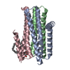
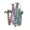
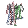
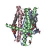
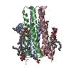
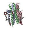
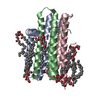
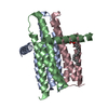
- PDB-4uxz: Structure of delta7-DgkA-syn in 7.9 MAG to 2.18 angstrom resolution -
+ Open data

- Basic information
| Entry | Database: PDB / ID: 4uxz | ||||||
|---|---|---|---|---|---|---|---|
| Title | Structure of delta7-DgkA-syn in 7.9 MAG to 2.18 angstrom resolution | ||||||
 Components Components | DIACYLGLYCEROL KINASE-DELTA 7 | ||||||
 Keywords Keywords |  TRANSFERASE / DIACYLGLYEROL KINASE / IN MESO CRYSTALLIZATION / LIPID CUBIC PHASE / LIPIDIC CUBIC PHASE / LIPID MESOPHASE / LIPIDIC MESOPHASE / MEMBRANE PROTEIN / MONOACYLGLYCEROL TRANSFERASE / DIACYLGLYEROL KINASE / IN MESO CRYSTALLIZATION / LIPID CUBIC PHASE / LIPIDIC CUBIC PHASE / LIPID MESOPHASE / LIPIDIC MESOPHASE / MEMBRANE PROTEIN / MONOACYLGLYCEROL | ||||||
| Function / homology |  Function and homology information Function and homology informationdiacylglycerol kinase (ATP) / ATP-dependent diacylglycerol kinase activity / phosphatidic acid biosynthetic process / response to UV / phosphorylation / ATP binding / membrane / identical protein binding / metal ion binding / plasma membraneSimilarity search - Function | ||||||
| Biological species |  ESCHERICHIA COLI K-12 (bacteria) ESCHERICHIA COLI K-12 (bacteria) | ||||||
| Method | X-RAY DIFFRACTION / SYNCHROTRON / MOLECULAR REPLACEMENT / Resolution: 2.18 Å | ||||||
 Authors Authors | Li, D. / Howe, N. / Caffrey, M. | ||||||
 Citation Citation | Journal: Nat.Commun. / Year: 2015 Title: Ternary Structure Reveals Mechanism of a Membrane Diacylglycerol Kinase. Authors: Li, D. / Stansfeld, P.J. / Sansom, M.S.P. / Keogh, A. / Vogeley, L. / Howe, N. / Lyons, J.A. / Aragao, D. / Fromme, P. / Fromme, R. / Basu, S. / Grotjohann, I. / Kupitz, C. / Rendek, K. / ...Authors: Li, D. / Stansfeld, P.J. / Sansom, M.S.P. / Keogh, A. / Vogeley, L. / Howe, N. / Lyons, J.A. / Aragao, D. / Fromme, P. / Fromme, R. / Basu, S. / Grotjohann, I. / Kupitz, C. / Rendek, K. / Weierstall, U. / Zatsepin, N.A. / Cherezov, V. / Liu, W. / Bandaru, S. / English, N.J. / Gati, C. / Barty, A. / Yefanov, O. / Chapman, H.N. / Diederichs, K. / Messerschmidt, M. / Boutet, S. / Williams, G.J. / Marvin Seibert, M. / Caffrey, M. | ||||||
| History |
|
- Structure visualization
| Structure viewer | Molecule: MolmilJmol/JSmol |
|---|
- Downloads & links
-Download
| PDBx/mmCIF format | 4uxz.cif.gz | 143.7 KB | Display | PDBx/mmCIF format |
|---|---|---|---|---|
| PDB format | pdb4uxz.ent.gz | 114.2 KB | Display | PDB format |
| PDBx/mmJSON format | 4uxz.json.gz | Tree view | PDBx/mmJSON format | |
| Others |  Other downloads Other downloads |
-Validation report
| Arichive directory | https://data.pdbj.org/pub/pdb/validation_reports/ux/4uxzftp://data.pdbj.org/pub/pdb/validation_reports/ux/4uxz | HTTPS FTP |
|---|
-Related structure data
| Related structure data |  4uxwC  4uxxC  4uyoC 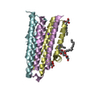 3ze3S C: citing same article ( S: Starting model for refinement |
|---|---|
| Similar structure data |


-Links
 PDBj
PDBj


- Assembly
| Deposited unit | 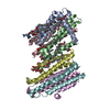
| ||||||||
|---|---|---|---|---|---|---|---|---|---|
| 1 | 
| ||||||||
| 2 | 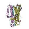
| ||||||||
| Unit cell |
|
-Components
-Protein , 1 types, 6 molecules ABCDEF
| #1: Protein | Mass: 14204.451 Da / Num. of mol.: 6 / Mutation: YES Source method: isolated from a genetically manipulated source Source: (gene. exp.) ESCHERICHIA COLI K-12 (bacteria) / Plasmid: PTRCHISB-DGKA-DELTA7 / Production host: ESCHERICHIA COLI (E. coli) / Strain (production host): WH1061 / References: UniProt: P0ABN1, diacylglycerol kinase (ATP) |
|---|
-Non-polymers , 6 types, 145 molecules 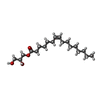


| #2: Chemical | ChemComp-79N / (  Mass: 328.487 Da / Num. of mol.: 4 / Source method: obtained synthetically / Formula: C19H36O4 Mass: 328.487 Da / Num. of mol.: 4 / Source method: obtained synthetically / Formula: C19H36O4#3: Chemical | ChemComp-79M / (  Mass: 328.487 Da / Num. of mol.: 7 / Source method: obtained synthetically / Formula: C19H36O4 Mass: 328.487 Da / Num. of mol.: 7 / Source method: obtained synthetically / Formula: C19H36O4#4: Chemical | ChemComp-ZN / |  Mass: 65.409 Da / Num. of mol.: 1 / Source method: obtained synthetically / Formula: Zn Mass: 65.409 Da / Num. of mol.: 1 / Source method: obtained synthetically / Formula: Zn#5: Chemical | ChemComp-FLC / | Citric acid Mass: 189.100 Da / Num. of mol.: 1 / Source method: obtained synthetically / Formula: C6H5O7 Mass: 189.100 Da / Num. of mol.: 1 / Source method: obtained synthetically / Formula: C6H5O7#6: Chemical | ChemComp-ACT / | Acetate Mass: 59.044 Da / Num. of mol.: 1 / Source method: obtained synthetically / Formula: C2H3O2 Mass: 59.044 Da / Num. of mol.: 1 / Source method: obtained synthetically / Formula: C2H3O2#7: Water | ChemComp-HOH / | WaterMass: 18.015 Da / Num. of mol.: 131 / Source method: isolated from a natural source / Formula: H2O |
|---|
-Details
| Sequence details | THE PROTEIN CONTAINS AN N-TERMINAL HIS TAG 'GHHHHHHEL'. COMPARED TO THE WILDTYPE FORM, THE PROTEIN ...THE PROTEIN CONTAINS AN N-TERMINAL HIS TAG 'GHHHHHHEL'. COMPARED TO THE WILDTYPE FORM, THE PROTEIN HAS SEVEN MUTATIONS. THEY ARE A41C, C46A, I53V, I70L, M96L, V107D AND C113A. |
|---|
-Experimental details
-Experiment
| Experiment | Method: X-RAY DIFFRACTION / Number of used crystals: 6 |
|---|
- Sample preparation
| Crystal | Density Matthews: 2.93 Å3/Da / Density % sol: 58.04 % / Description: NONE |
|---|---|
| Crystal grow | Temperature: 277 K / Method: lipidic cubic phase / pH: 5.6 Details: 4-6 %(V/V) 2-METHYL-2, 4-PENTANEDIOL (MPD), 0.1 M SODIUM CHLORIDE, 0.06 M MAGNESIUM ACETATE, 0.1 M SODIUM CITRATE/HCL PH 5.6. CRYSTALLIZED USING THE IN MESO (LIPID CUBIC PHASE) METHOD AT 4 ...Details: 4-6 %(V/V) 2-METHYL-2, 4-PENTANEDIOL (MPD), 0.1 M SODIUM CHLORIDE, 0.06 M MAGNESIUM ACETATE, 0.1 M SODIUM CITRATE/HCL PH 5.6. CRYSTALLIZED USING THE IN MESO (LIPID CUBIC PHASE) METHOD AT 4 DEGREES CELCIUS WITH THE 7.9 MONOACYLGLYCEROL (7.9 MAG) AS THE HOSTING LIPID. |
-Data collection
| Diffraction | Mean temperature: 100 K |
|---|---|
| Diffraction source | Source: SYNCHROTRON / Site: SLS  / Beamline: X06DA / Wavelength: 1.03315 / Beamline: X06DA / Wavelength: 1.03315 |
| Detector | Type: DECTRIS PILATUS 6M / Detector: PIXEL / Date: Feb 6, 2013 / Details: MIRRORS |
| Radiation | Monochromator: DOUBLE CRYSTAL / Protocol: SINGLE WAVELENGTH / Monochromatic (M) / Laue (L): M / Scattering type: x-ray |
| Radiation wavelength | Wavelength: 1.03315 Å / Relative weight: 1 |
| Reflection | Resolution: 2.18→75.02 Å / Num. obs: 51820 / % possible obs: 99.1 % / Observed criterion σ(I): -3 / Redundancy: 6.7 % / Biso Wilson estimate: 42.36 Å2 / Rmerge(I) obs: 0.1 / Net I/σ(I): 12.1 |
| Reflection shell | Resolution: 2.18→2.24 Å / Redundancy: 6.4 % / Rmerge(I) obs: 1.27 / Mean I/σ(I) obs: 1.7 / % possible all: 96.8 |
- Processing
| Software |
| |||||||||||||||||||||||||||||||||||||||||||||||||||||||||||||||||||||||||||||||||||||||||||||||||||||||||||||||||||||||||||||||||||||
|---|---|---|---|---|---|---|---|---|---|---|---|---|---|---|---|---|---|---|---|---|---|---|---|---|---|---|---|---|---|---|---|---|---|---|---|---|---|---|---|---|---|---|---|---|---|---|---|---|---|---|---|---|---|---|---|---|---|---|---|---|---|---|---|---|---|---|---|---|---|---|---|---|---|---|---|---|---|---|---|---|---|---|---|---|---|---|---|---|---|---|---|---|---|---|---|---|---|---|---|---|---|---|---|---|---|---|---|---|---|---|---|---|---|---|---|---|---|---|---|---|---|---|---|---|---|---|---|---|---|---|---|---|---|---|
| Refinement | Method to determine structure: MOLECULAR REPLACEMENT Starting model: PDB ENTRY 3ZE3 Resolution: 2.18→57.965 Å / SU ML: 0.27 / σ(F): 1.34 / Phase error: 24.15 / Stereochemistry target values: ML Details: THERE ARE SIX NCS-RELATED MOLECULES IN THE ASYMMETRIC UNIT BUT NCS RESTRAINTS WERE NOT USED IN THE REFINEMENT.
| |||||||||||||||||||||||||||||||||||||||||||||||||||||||||||||||||||||||||||||||||||||||||||||||||||||||||||||||||||||||||||||||||||||
| Solvent computation | Shrinkage radii: 0.9 Å / VDW probe radii: 1.11 Å / Solvent model: FLAT BULK SOLVENT MODEL | |||||||||||||||||||||||||||||||||||||||||||||||||||||||||||||||||||||||||||||||||||||||||||||||||||||||||||||||||||||||||||||||||||||
| Displacement parameters | Biso mean: 60.07 Å2 | |||||||||||||||||||||||||||||||||||||||||||||||||||||||||||||||||||||||||||||||||||||||||||||||||||||||||||||||||||||||||||||||||||||
| Refinement step | Cycle: LAST / Resolution: 2.18→57.965 Å
| |||||||||||||||||||||||||||||||||||||||||||||||||||||||||||||||||||||||||||||||||||||||||||||||||||||||||||||||||||||||||||||||||||||
| Refine LS restraints |
| |||||||||||||||||||||||||||||||||||||||||||||||||||||||||||||||||||||||||||||||||||||||||||||||||||||||||||||||||||||||||||||||||||||
| LS refinement shell |
|
