[English] 日本語
Yorodumi
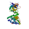
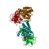
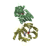
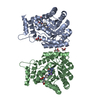
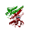
- PDB-4uxl: Structure of Human ROS1 Kinase Domain in Complex with PF-06463922 -

+
Open data


ID or keywords:

Loading...

-
Basic information

Entry
Database: PDB / ID: 4uxl
TitleStructure of Human ROS1 Kinase Domain in Complex with PF-06463922
ComponentsPROTO-ONCOGENE TYROSINE-PROTEIN KINASE ROS
KeywordsTRANSFERASE / INHIBITOR / ROS
Function / homology
Function and homology information


columnar/cuboidal epithelial cell development / regulation of TOR signaling / transmembrane receptor protein tyrosine kinase activity / regulation of ERK1 and ERK2 cascade / regulation of cell growth / receptor protein-tyrosine kinase / cell surface receptor protein tyrosine kinase signaling pathway / spermatogenesis / protein phosphatase binding / protein tyrosine kinase activity ...columnar/cuboidal epithelial cell development / regulation of TOR signaling / transmembrane receptor protein tyrosine kinase activity / regulation of ERK1 and ERK2 cascade / regulation of cell growth / receptor protein-tyrosine kinase / cell surface receptor protein tyrosine kinase signaling pathway / spermatogenesis / protein phosphatase binding / protein tyrosine kinase activity / cell differentiation / receptor complex / protein phosphorylation / ATP binding / membrane / plasma membrane
Similarity search - Function
LDLR class B repeat / Low-density lipoprotein-receptor YWTD domain / Tyrosine-protein kinase, receptor class II, conserved site / Six-bladed beta-propeller, TolB-like / Receptor tyrosine kinase class II signature. / Fibronectin type III domain / Fibronectin type 3 domain / Fibronectin type-III domain profile. / Fibronectin type III / Fibronectin type III superfamily ...LDLR class B repeat / Low-density lipoprotein-receptor YWTD domain / Tyrosine-protein kinase, receptor class II, conserved site / Six-bladed beta-propeller, TolB-like / Receptor tyrosine kinase class II signature. / Fibronectin type III domain / Fibronectin type 3 domain / Fibronectin type-III domain profile. / Fibronectin type III / Fibronectin type III superfamily / Tyrosine-protein kinase, catalytic domain / Tyrosine kinase, catalytic domain / Tyrosine protein kinases specific active-site signature. / Tyrosine-protein kinase, active site / Protein tyrosine and serine/threonine kinase / Serine-threonine/tyrosine-protein kinase, catalytic domain / Transferase(Phosphotransferase) domain 1 / Transferase(Phosphotransferase); domain 1 / Phosphorylase Kinase; domain 1 / Phosphorylase Kinase; domain 1 / Protein kinase, ATP binding site / Protein kinases ATP-binding region signature. / Immunoglobulin-like fold / Protein kinase domain profile. / Protein kinase domain / Protein kinase-like domain superfamily / 2-Layer Sandwich / Orthogonal Bundle / Mainly Alpha / Alpha Beta
Similarity search - Domain/homology
Chem-5P8 / Proto-oncogene tyrosine-protein kinase ROS
Similarity search - Component
Biological speciesHOMO SAPIENS (human)
MethodX-RAY DIFFRACTION / SYNCHROTRON / MOLECULAR REPLACEMENT / Resolution: 2.4 Å
AuthorsMcTigue, M. / Deng, Y. / Liu, W. / Brooun, A. / Stewart, A.
CitationJournal: Proc.Natl.Acad.Sci.USA / Year: 2015
Title: Pf-06463922 is a Potent and Selective Next-Generation Ros1/Alk Inhibitor Capable of Blocking Crizotinib-Resistant Ros1 Mutations.
Authors: Zou, H.Y. / Li, Q. / Engstrom, L.D. / West, M. / Appleman, V. / Wong, K.A. / Mctigue, M. / Deng, Y. / Liu, W. / Brooun, A. / Timofeevski, S. / Mcdonnell, S.R.P. / Jiang, P. / Falk, M.D. / ...Authors: Zou, H.Y. / Li, Q. / Engstrom, L.D. / West, M. / Appleman, V. / Wong, K.A. / Mctigue, M. / Deng, Y. / Liu, W. / Brooun, A. / Timofeevski, S. / Mcdonnell, S.R.P. / Jiang, P. / Falk, M.D. / Lappin, P.B. / Affolter, T. / Nichols, T. / Hu, W. / Lam, J. / Johnson, T.W. / Smeal, T. / Charest, A. / Fantin, V.R.
History
DepositionAug 25, 2014Deposition site: PDBE / Processing site: PDBE
Revision 1.0Mar 11, 2015Provider: repository / Type: Initial release
Revision 1.1Mar 18, 2015Group: Database references
Revision 1.2Apr 15, 2015Group: Database references
Revision 1.3Apr 24, 2019Group: Data collection / Other / Source and taxonomy
Category: entity_src_gen / pdbx_database_proc / pdbx_database_status
Item: _entity_src_gen.pdbx_host_org_cell_line / _pdbx_database_status.recvd_author_approval
Revision 1.4May 8, 2019Group: Data collection / Experimental preparation
Category: database_PDB_rev / database_PDB_rev_record / exptl_crystal_grow
Item: _exptl_crystal_grow.method / _exptl_crystal_grow.temp
Revision 1.5Jan 10, 2024Group: Data collection / Database references ...Data collection / Database references / Derived calculations / Other / Refinement description
Category: chem_comp_atom / chem_comp_bond ...chem_comp_atom / chem_comp_bond / database_2 / pdbx_database_status / pdbx_initial_refinement_model / software / struct_site
Item: _database_2.pdbx_DOI / _database_2.pdbx_database_accession ..._database_2.pdbx_DOI / _database_2.pdbx_database_accession / _pdbx_database_status.status_code_sf / _software.name / _struct_site.pdbx_auth_asym_id / _struct_site.pdbx_auth_comp_id / _struct_site.pdbx_auth_seq_id

-
Structure visualization

Structure viewerMolecule:
MolmilJmol/JSmol

Downloads & links

-
Assembly

Deposited unit
A: PROTO-ONCOGENE TYROSINE-PROTEIN KINASE ROS
hetero molecules


Theoretical massNumber of molelcules
Total (without water)37,4212
Polymers37,0141
Non-polymers4061
Water2,450136
1
A: PROTO-ONCOGENE TYROSINE-PROTEIN KINASE ROS
hetero molecules

A: PROTO-ONCOGENE TYROSINE-PROTEIN KINASE ROS
hetero molecules


Theoretical massNumber of molelcules
Total (without water)74,8424
Polymers74,0292
Non-polymers8132
Water362
TypeNameSymmetry operationNumber
identity operation1_555x,y,z1
crystal symmetry operation4_655-x+1,-y,z1
Buried area3830 Å2
ΔGint-20.3 kcal/mol
Surface area26520 Å2
MethodPISA
Unit cell
Length a, b, c (Å)105.844, 105.844, 49.843
Angle α, β, γ (deg.)90.00, 90.00, 120.00
Int Tables number172
Space group name H-MP64

-
Components

#1: Protein PROTO-ONCOGENE TYROSINE-PROTEIN KINASE ROS / PROTO-ONCOGENE C-ROS / PROTO-ONCOGENE C-ROS-1 / RECEPTOR TYROSINE KINASE C-ROS ONCOGENE 1 / C-ROS ...PROTO-ONCOGENE C-ROS / PROTO-ONCOGENE C-ROS-1 / RECEPTOR TYROSINE KINASE C-ROS ONCOGENE 1 / C-ROS RECEPTOR TYROSINE KINASE


Mass: 37014.488 Da / Num. of mol.: 1 / Fragment: KINASE DOMAIN, UNP RESIDUES 1934-2232
Source method: isolated from a genetically manipulated source
Source: (gene. exp.) HOMO SAPIENS (human) / Plasmid: PFASTBAC / Cell line (production host): Sf21 / Production host: SPODOPTERA FRUGIPERDA (fall armyworm)
References: UniProt: P08922, receptor protein-tyrosine kinase
#2: Chemical ChemComp-5P8 / (10R)-7-amino-12-fluoro-2,10,16-trimethyl-15-oxo-10,15,16,17-tetrahydro-2H-8,4-(metheno)pyrazolo[4,3-h][2,5,11]benzoxadiazacyclotetradecine-3-carbonitrile / Lorlatinib


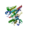
Mass: 406.413 Da / Num. of mol.: 1 / Source method: obtained synthetically / Formula: C21H19FN6O2 / Comment: anticancer, inhibitor*YM
#3: Water ChemComp-HOH / water / Water


Mass: 18.015 Da / Num. of mol.: 136 / Source method: isolated from a natural source / Formula: H2O
Nonpolymer details(10R)-7-AMINO-12-FLUORO-2,10,16-TRIMETHYL-15-OXO-10,15,16, 17-TETRAHYDRO-2H-8,4-(METHENO) ...(10R)-7-AMINO-12-FLUORO-2,10,16-TRIMETHYL-15-OXO-10,15,16, 17-TETRAHYDRO-2H-8,4-(METHENO)PYRAZOLO(4,3-H)(2,5, 11)BENZOXADIAZACYCLOTETRADECINE-3-CARBONITRILE) (UNL): HET IS THE SAME AS 5P8 IN ENTRY 4CLJ

-
Experimental details

-
Experiment

ExperimentMethod: X-RAY DIFFRACTION / Number of used crystals: 1

-
Sample preparation

CrystalDensity Matthews: 2.7 Å3/Da / Density % sol: 55 % / Description: NONE
Crystal growTemperature: 286 K / Method: vapor diffusion, hanging drop / pH: 5.6
Details: CRYSTALS WERE OBTAINED BY THE HANGING DROP VAPOR DIFFUSION METHOD AT 13 DEGREES C BY MIXING 1.5 MICROLITERS OF SOLUTION CONTAINING A 1:3 MOLAR RATIO OF PHOSPHORYLATED ROS1 KD (12.2MG/ML) TO ...Details: CRYSTALS WERE OBTAINED BY THE HANGING DROP VAPOR DIFFUSION METHOD AT 13 DEGREES C BY MIXING 1.5 MICROLITERS OF SOLUTION CONTAINING A 1:3 MOLAR RATIO OF PHOSPHORYLATED ROS1 KD (12.2MG/ML) TO PF-06463922 AND 1.5 MICROLITERS OF RESERVOIR SOLUTION CONTAINING 25% (W/V) PEG 3350, 0.6M POTASSIUM THIOCYANATE AND 0.1M SODIUM CITRATE TRIBASIC DIHYDRATE, PH5.6. CRYSTALS WERE FLASH-FROZEN IN LIQUID NITROGEN AFTER TRANSFER TO 2 MICROLITERS OF RESERVOIR SOLUTION CONTAINING 25% (V/V) GLYCEROL AS A CRYOPROTECTANT.

-
Data collection

DiffractionMean temperature: 87 K
Diffraction sourceSource: SYNCHROTRON / Site: APS / Beamline: 17-ID / Wavelength: 1
DetectorType: DECTRIS PILATUS 6M / Detector: PIXEL / Date: Apr 19, 2012
RadiationProtocol: SINGLE WAVELENGTH / Monochromatic (M) / Laue (L): M / Scattering type: x-ray
Radiation wavelengthWavelength: 1 Å / Relative weight: 1
ReflectionResolution: 2.21→91.66 Å / Num. obs: 15684 / % possible obs: 97.1 % / Observed criterion σ(I): 2 / Redundancy: 6.7 % / Biso Wilson estimate: 41.4 Å2 / Rmerge(I) obs: 0.04 / Net I/σ(I): 27
Reflection shellResolution: 2.21→2.33 Å / Redundancy: 4.4 % / Rmerge(I) obs: 0.42 / Mean I/σ(I) obs: 2.8 / % possible all: 62.1

-
Processing

Software
NameVersionClassification
CNS2005refinement
autoPROCdata reduction
SCALAdata scaling
CNXphasing
RefinementMethod to determine structure: MOLECULAR REPLACEMENT
Starting model: PDB ENTRY 3ZBF
Resolution: 2.4→91.66 Å / Rfactor Rfree error: 0.014 / Data cutoff high absF: 1439110.98 / Data cutoff low absF: 0 / Isotropic thermal model: RESTRAINED / Cross valid method: THROUGHOUT / σ(F): 0
RfactorNum. reflection% reflectionSelection details
Rfree0.281 379 3 %RANDOM
Rwork0.211 ---
obs0.211 12648 100 %-
Solvent computationSolvent model: FLAT MODEL / Bsol: 55.3806 Å2 / ksol: 0.360053 e/Å3
Displacement parametersBiso mean: 50.4 Å2
Baniso -1Baniso -2Baniso -3
1--5.98 Å22.43 Å20 Å2
2---5.98 Å20 Å2
3---11.97 Å2
Refine analyze
FreeObs
Luzzati coordinate error0.38 Å0.27 Å
Luzzati d res low-5 Å
Luzzati sigma a0.28 Å0.17 Å
Refinement stepCycle: LAST / Resolution: 2.4→91.66 Å
ProteinNucleic acidLigandSolventTotal
Num. atoms2310 0 30 136 2476
Refine LS restraints
Refine-IDTypeDev idealDev ideal target
X-RAY DIFFRACTIONc_bond_d0.006
X-RAY DIFFRACTIONc_bond_d_na
X-RAY DIFFRACTIONc_bond_d_prot
X-RAY DIFFRACTIONc_angle_d
X-RAY DIFFRACTIONc_angle_d_na
X-RAY DIFFRACTIONc_angle_d_prot
X-RAY DIFFRACTIONc_angle_deg0.9
X-RAY DIFFRACTIONc_angle_deg_na
X-RAY DIFFRACTIONc_angle_deg_prot
X-RAY DIFFRACTIONc_dihedral_angle_d21.8
X-RAY DIFFRACTIONc_dihedral_angle_d_na
X-RAY DIFFRACTIONc_dihedral_angle_d_prot
X-RAY DIFFRACTIONc_improper_angle_d0.62
X-RAY DIFFRACTIONc_improper_angle_d_na
X-RAY DIFFRACTIONc_improper_angle_d_prot
X-RAY DIFFRACTIONc_mcbond_it1.661.5
X-RAY DIFFRACTIONc_mcangle_it2.822
X-RAY DIFFRACTIONc_scbond_it2.132
X-RAY DIFFRACTIONc_scangle_it3.112.5
Refine LS restraints NCSNCS model details: NONE
LS refinement shellResolution: 2.4→2.55 Å / Rfactor Rfree error: 0.046 / Total num. of bins used: 6
RfactorNum. reflection% reflection
Rfree0.37 65 3.1 %
Rwork0.241 2024 -
obs--100 %
Xplor file
Refine-IDSerial noParam fileTopol file
X-RAY DIFFRACTION1PROTEIN_REP.PARAMPROTEIN.TOP
X-RAY DIFFRACTION2DNA-RNA_REP.PARAMDNA-RNA.TOP
X-RAY DIFFRACTION3WATER_REP.PARAMWATER.TOP
X-RAY DIFFRACTION4ION.PARAMION.TOP
X-RAY DIFFRACTION5UNL.PARAMUNL.TOP

+
About Yorodumi

-
News

-
Feb 9, 2022. New format data for meta-information of EMDB entries

New format data for meta-information of EMDB entries

  • Version 3 of the EMDB header file is now the official format.
  • The previous official version 1.9 will be removed from the archive.

Related info.:EMDB header

External links:wwPDB to switch to version 3 of the EMDB data model

-
Aug 12, 2020. Covid-19 info

Covid-19 info

URL: https://pdbj.org/emnavi/covid19.php

New page: Covid-19 featured information page in EM Navigator.

Related info.:Covid-19 info / Mar 5, 2020. Novel coronavirus structure data

+
Mar 5, 2020. Novel coronavirus structure data

Novel coronavirus structure data

Related info.:Yorodumi Speices / Aug 12, 2020. Covid-19 info

External links:COVID-19 featured content - PDBj / Molecule of the Month (242):Coronavirus Proteases

+
Jan 31, 2019. EMDB accession codes are about to change! (news from PDBe EMDB page)

EMDB accession codes are about to change! (news from PDBe EMDB page)

  • The allocation of 4 digits for EMDB accession codes will soon come to an end. Whilst these codes will remain in use, new EMDB accession codes will include an additional digit and will expand incrementally as the available range of codes is exhausted. The current 4-digit format prefixed with “EMD-” (i.e. EMD-XXXX) will advance to a 5-digit format (i.e. EMD-XXXXX), and so on. It is currently estimated that the 4-digit codes will be depleted around Spring 2019, at which point the 5-digit format will come into force.
  • The EM Navigator/Yorodumi systems omit the EMD- prefix.

Related info.:Q: What is EMD? / ID/Accession-code notation in Yorodumi/EM Navigator

External links:EMDB Accession Codes are Changing Soon! / Contact to PDBj

+
Jul 12, 2017. Major update of PDB

Major update of PDB

  • wwPDB released updated PDB data conforming to the new PDBx/mmCIF dictionary.
  • This is a major update changing the version number from 4 to 5, and with Remediation, in which all the entries are updated.
  • In this update, many items about electron microscopy experimental information are reorganized (e.g. em_software).
  • Now, EM Navigator and Yorodumi are based on the updated data.

External links:wwPDB Remediation / Enriched Model Files Conforming to OneDep Data Standards Now Available in the PDB FTP Archive

-
Yorodumi

Thousand views of thousand structures

  • Yorodumi is a browser for structure data from EMDB, PDB, SASBDB, etc.
  • This page is also the successor to EM Navigator detail page, and also detail information page/front-end page for Omokage search.
  • The word "yorodu" (or yorozu) is an old Japanese word meaning "ten thousand". "mi" (miru) is to see.

Related info.:EMDB / PDB / SASBDB / Comparison of 3 databanks / Yorodumi Search / Aug 31, 2016. New EM Navigator & Yorodumi / Yorodumi Papers / Jmol/JSmol / Function and homology information / Changes in new EM Navigator and Yorodumi

Read more