 Movie
Movie Controller


+ Open data

- Basic information
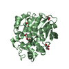
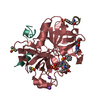
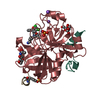
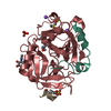
| Entry | Database: PDB / ID: 4ue7 | |||||||||
|---|---|---|---|---|---|---|---|---|---|---|
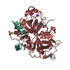
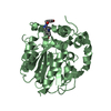
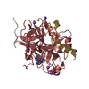
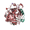
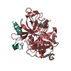
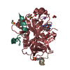
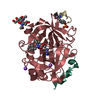
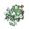
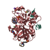
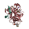
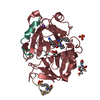
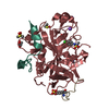
| Title | Thrombin in complex with 1-amidinopiperidine | |||||||||
 Components Components |
| |||||||||
 Keywords Keywords |  HYDROLASE / HYDROLASE INHIBITOR COMPLEX / SERINE PROTEASE / BLOOD COAGULATION / BLOOD CLOTTING / CONVERTION OF FIBRINOGEN TO FIBRIN / BLOOD CLOTTING INHIBITOR / THROMBIN INHIBITOR / GLYCOSYLATION / BLOOD HYDROLASE / HYDROLASE INHIBITOR COMPLEX / SERINE PROTEASE / BLOOD COAGULATION / BLOOD CLOTTING / CONVERTION OF FIBRINOGEN TO FIBRIN / BLOOD CLOTTING INHIBITOR / THROMBIN INHIBITOR / GLYCOSYLATION / BLOOD | |||||||||
| Function / homology |  Function and homology information Function and homology informationpositive regulation of lipid kinase activity / positive regulation of phospholipase C-activating G protein-coupled receptor signaling pathway / cytolysis by host of symbiont cells / thrombospondin receptor activity / Defective factor XII causes hereditary angioedema / thrombin / neutrophil-mediated killing of gram-negative bacterium / regulation of blood coagulation / ligand-gated ion channel signaling pathway / Defective F8 cleavage by thrombin ...positive regulation of lipid kinase activity / positive regulation of phospholipase C-activating G protein-coupled receptor signaling pathway / cytolysis by host of symbiont cells / thrombospondin receptor activity / Defective factor XII causes hereditary angioedema / thrombin / neutrophil-mediated killing of gram-negative bacterium / regulation of blood coagulation / ligand-gated ion channel signaling pathway / Defective F8 cleavage by thrombin / Platelet Aggregation (Plug Formation) / negative regulation of platelet activation / negative regulation of astrocyte differentiation / negative regulation of cytokine production involved in inflammatory response / positive regulation of collagen biosynthetic process / positive regulation of blood coagulation / negative regulation of fibrinolysis / Gamma-carboxylation of protein precursors / Transport of gamma-carboxylated protein precursors from the endoplasmic reticulum to the Golgi apparatus / Common Pathway of Fibrin Clot Formation / Removal of aminoterminal propeptides from gamma-carboxylated proteins / regulation of cytosolic calcium ion concentration / fibrinolysis / Intrinsic Pathway of Fibrin Clot Formation / Peptide ligand-binding receptors / positive regulation of release of sequestered calcium ion into cytosol / Regulation of Complement cascade / acute-phase response / Cell surface interactions at the vascular wall / lipopolysaccharide binding / negative regulation of proteolysis / positive regulation of receptor signaling pathway via JAK-STAT / growth factor activity / serine-type endopeptidase inhibitor activity / positive regulation of insulin secretion / platelet activation / response to wounding / Golgi lumen / positive regulation of protein localization to nucleus / Regulation of Insulin-like Growth Factor (IGF) transport and uptake by Insulin-like Growth Factor Binding Proteins (IGFBPs) / positive regulation of reactive oxygen species metabolic process / blood coagulation / antimicrobial humoral immune response mediated by antimicrobial peptide / Thrombin signalling through proteinase activated receptors (PARs) / heparin binding / regulation of cell shape / positive regulation of cell growth / G alpha (q) signalling events / collagen-containing extracellular matrix / blood microparticle / cell surface receptor signaling pathway / positive regulation of phosphatidylinositol 3-kinase/protein kinase B signal transduction / positive regulation of protein phosphorylation / G protein-coupled receptor signaling pathway / endoplasmic reticulum lumen / signaling receptor binding / serine-type endopeptidase activity / calcium ion binding / positive regulation of cell population proliferation / proteolysis / extracellular space / extracellular exosome / extracellular region / plasma membraneSimilarity search - Function | |||||||||
| Biological species |  HOMO SAPIENS (human) HOMO SAPIENS (human) HIRUDO MEDICINALIS (medicinal leech) HIRUDO MEDICINALIS (medicinal leech) | |||||||||
| Method | X-RAY DIFFRACTION / SYNCHROTRON / MOLECULAR REPLACEMENT / Resolution: 1.129 Å | |||||||||
 Authors Authors | Ruehmann, E. / Heine, A. / Klebe, G. | |||||||||
 Citation Citation | Journal: J.Med.Chem. / Year: 2015 Title: Fragments Can Bind Either More Enthalpy or Entropy-Driven: Crystal Structures and Residual Hydration Pattern Suggest Why. Authors: Ruehmann, E. / Betz, M. / Heine, A. / Klebe, G. | |||||||||
| History |
| |||||||||
| Remark 700 | SHEET DETERMINATION METHOD: DSSP THE SHEETS PRESENTED AS "HB" IN EACH CHAIN ON SHEET RECORDS BELOW ... SHEET DETERMINATION METHOD: DSSP THE SHEETS PRESENTED AS "HB" IN EACH CHAIN ON SHEET RECORDS BELOW IS ACTUALLY AN 6-STRANDED BARREL THIS IS REPRESENTED BY A 7-STRANDED SHEET IN WHICH THE FIRST AND LAST STRANDS ARE IDENTICAL. |
- Structure visualization
| Structure viewer | Molecule: MolmilJmol/JSmol |
|---|
- Downloads & links
-Download
| PDBx/mmCIF format | 4ue7.cif.gz | 204.1 KB | Display | PDBx/mmCIF format |
|---|---|---|---|---|
| PDB format | pdb4ue7.ent.gz | 164.7 KB | Display | PDB format |
| PDBx/mmJSON format | 4ue7.json.gz | Tree view | PDBx/mmJSON format | |
| Others |  Other downloads Other downloads |
-Validation report
| Arichive directory | https://data.pdbj.org/pub/pdb/validation_reports/ue/4ue7ftp://data.pdbj.org/pub/pdb/validation_reports/ue/4ue7 | HTTPS FTP |
|---|
-Related structure data
| Related structure data |  4ud9C 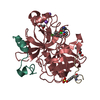 4udwC 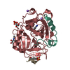 4uehC 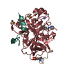 5af9C  5afyC 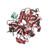 5afzC  5ahgC  1h8dS S: Starting model for refinement C: citing same article ( |
|---|---|
| Similar structure data |


-Links
 PDBj
PDBj


- Assembly
| Deposited unit | 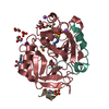
| |||||||||
|---|---|---|---|---|---|---|---|---|---|---|
| 1 |
| |||||||||
| Unit cell |
| |||||||||
| Components on special symmetry positions |
|

-Components
-Protein/peptide , 2 types, 2 molecules IL
| #2: Protein/peptide | Mass: 1548.580 Da / Num. of mol.: 1 / Fragment: UNP RESIDUES 61-72 / Source method: obtained synthetically / Details: HIRUDIN (54-65) (SULFATED) / Source: (synth.) HIRUDO MEDICINALIS (medicinal leech) / References: UniProt: P09945 |
|---|---|
| #3: Protein/peptide | / PROTHROMBIN / COAGULATION FACTOR II / ACTIVATION PEPTIDE FRAGMENT 1 / ACTIVATION PEPTIDE FRAGMENT 2 Mass: 3317.741 Da / Num. of mol.: 1 / Fragment: THROMBIN LIGHT CHAIN, UNP RESIDUES 333-360 / Source method: isolated from a natural source / Details: PURIFIED FROM HUMAN BLOOD PLASMA / Source: (natural) HOMO SAPIENS (human) / References: UniProt: P00734, thrombin |
-Protein / Sugars , 2 types, 2 molecules H
| #11: Sugar | ChemComp-NAG / N-Acetylglucosamine Type: D-saccharide, beta linking / Mass: 221.208 Da / Num. of mol.: 1 Type: D-saccharide, beta linking / Mass: 221.208 Da / Num. of mol.: 1Source method: isolated from a genetically manipulated source Formula: C8H15NO6 |
|---|---|
| #1: Protein | / PROTHROMBIN / COAGULATION FACTOR II / ACTIVATION PEPTIDE FRAGMENT 1 / ACTIVATION PEPTIDE FRAGMENT 2 ...PROTHROMBIN / COAGULATION FACTOR II / ACTIVATION PEPTIDE FRAGMENT 1 / ACTIVATION PEPTIDE FRAGMENT 2 / THROMBIN LIGHT CHAIN / THROMBIN HEAVY CHAIN Mass: 29651.105 Da / Num. of mol.: 1 / Fragment: THROMBIN HEAVY CHAIN, UNP RESIDUES 364-621 / Source method: isolated from a natural source / Details: PURIFIED FROM HUMAN BLOOD PLASMA / Source: (natural) HOMO SAPIENS (human) / References: UniProt: P00734, thrombin |
-Non-polymers , 8 types, 334 molecules 


| #4: Chemical | ChemComp-IOD / Iodide Mass: 126.904 Da / Num. of mol.: 1 / Source method: obtained synthetically / Formula: I Mass: 126.904 Da / Num. of mol.: 1 / Source method: obtained synthetically / Formula: I | ||||||||||||
|---|---|---|---|---|---|---|---|---|---|---|---|---|---|
| #5: Chemical | Glycerol Mass: 92.094 Da / Num. of mol.: 3 / Source method: obtained synthetically / Formula: C3H8O3 Mass: 92.094 Da / Num. of mol.: 3 / Source method: obtained synthetically / Formula: C3H8O3#6: Chemical |  Mass: 22.990 Da / Num. of mol.: 2 / Source method: obtained synthetically / Formula: Na Mass: 22.990 Da / Num. of mol.: 2 / Source method: obtained synthetically / Formula: Na#7: Chemical | ChemComp-PO4 / | Phosphate Mass: 94.971 Da / Num. of mol.: 1 / Source method: obtained synthetically / Formula: PO4 Mass: 94.971 Da / Num. of mol.: 1 / Source method: obtained synthetically / Formula: PO4#8: Chemical | Dimethyl sulfoxide Mass: 78.133 Da / Num. of mol.: 3 / Source method: obtained synthetically / Formula: C2H6OS / Comment: DMSO, precipitant*YM Mass: 78.133 Da / Num. of mol.: 3 / Source method: obtained synthetically / Formula: C2H6OS / Comment: DMSO, precipitant*YM#9: Chemical | ChemComp-EDO / | Ethylene glycol Mass: 62.068 Da / Num. of mol.: 1 / Source method: obtained synthetically / Formula: C2H6O2 Mass: 62.068 Da / Num. of mol.: 1 / Source method: obtained synthetically / Formula: C2H6O2#10: Chemical | ChemComp-MRZ / |  Mass: 127.188 Da / Num. of mol.: 1 / Source method: obtained synthetically / Formula: C6H13N3 Mass: 127.188 Da / Num. of mol.: 1 / Source method: obtained synthetically / Formula: C6H13N3#12: Water | ChemComp-HOH / | WaterMass: 18.015 Da / Num. of mol.: 322 / Source method: isolated from a natural source / Formula: H2O |
-Details
| Sequence details | ASP 14L WAS NOT BUILD DUE TO LACK OF ELECTRON DESITY RESIDUES 148-149E WERE NOT BUILD DUE TO LACK ...ASP 14L WAS NOT BUILD DUE TO LACK OF ELECTRON DESITY RESIDUES 148-149E WERE NOT BUILD DUE TO LACK OF ELECTRON DENSITY |
|---|
-Experimental details
-Experiment
| Experiment | Method: X-RAY DIFFRACTION / Number of used crystals: 1 |
|---|
- Sample preparation
| Crystal | Density Matthews: 2.48 Å3/Da / Density % sol: 50.49 % / Description: NONE |
|---|---|
| Crystal grow | pH: 7.5 Details: SEE MATERIALS AND METHODS SECTION OF PUBLICATION, pH 7.5 |
-Data collection
| Diffraction | Mean temperature: 113 K |
|---|---|
| Diffraction source | Source: SYNCHROTRON / Site: BESSY  / Beamline: 14.2 / Wavelength: 0.91841 / Beamline: 14.2 / Wavelength: 0.91841 |
| Detector | Type: MARMOSAIC 225 mm CCD / Detector: CCD / Date: Jul 18, 2013 |
| Radiation | Protocol: SINGLE WAVELENGTH / Monochromatic (M) / Laue (L): M / Scattering type: x-ray |
| Radiation wavelength | Wavelength: 0.91841 Å / Relative weight: 1 |
| Reflection | Resolution: 1.13→19.56 Å / Num. obs: 130988 / % possible obs: 99.5 % / Observed criterion σ(I): 2.5 / Redundancy: 3 % / Biso Wilson estimate: 10.67 Å2 / Rmerge(I) obs: 0.04 / Net I/σ(I): 24.5 |
| Reflection shell | Resolution: 1.13→1.15 Å / Redundancy: 2.7 % / Rmerge(I) obs: 0.39 / Mean I/σ(I) obs: 2.54 / % possible all: 99.5 |
- Processing
| Software |
| |||||||||||||||||||||||||||||||||||||||||||||||||||||||||||||||||||||||||||||||||||||||||||||||||||||||||||||||||||||||||||||||||||||||||||||||||||||||||||||||||||||||||||||||||||||||||||||||||||||||||||||||||||||||||
|---|---|---|---|---|---|---|---|---|---|---|---|---|---|---|---|---|---|---|---|---|---|---|---|---|---|---|---|---|---|---|---|---|---|---|---|---|---|---|---|---|---|---|---|---|---|---|---|---|---|---|---|---|---|---|---|---|---|---|---|---|---|---|---|---|---|---|---|---|---|---|---|---|---|---|---|---|---|---|---|---|---|---|---|---|---|---|---|---|---|---|---|---|---|---|---|---|---|---|---|---|---|---|---|---|---|---|---|---|---|---|---|---|---|---|---|---|---|---|---|---|---|---|---|---|---|---|---|---|---|---|---|---|---|---|---|---|---|---|---|---|---|---|---|---|---|---|---|---|---|---|---|---|---|---|---|---|---|---|---|---|---|---|---|---|---|---|---|---|---|---|---|---|---|---|---|---|---|---|---|---|---|---|---|---|---|---|---|---|---|---|---|---|---|---|---|---|---|---|---|---|---|---|---|---|---|---|---|---|---|---|---|---|---|---|---|---|---|---|
| Refinement | Method to determine structure: MOLECULAR REPLACEMENT Starting model: PDB ENTRY 1H8D Resolution: 1.129→19.563 Å / SU ML: 0.07 / σ(F): 1.34 / Phase error: 12.09 / Stereochemistry target values: ML / Details: RESIDUES 148-149E ARE DISORDERED
| |||||||||||||||||||||||||||||||||||||||||||||||||||||||||||||||||||||||||||||||||||||||||||||||||||||||||||||||||||||||||||||||||||||||||||||||||||||||||||||||||||||||||||||||||||||||||||||||||||||||||||||||||||||||||
| Solvent computation | Shrinkage radii: 0.9 Å / VDW probe radii: 1.11 Å / Solvent model: FLAT BULK SOLVENT MODEL | |||||||||||||||||||||||||||||||||||||||||||||||||||||||||||||||||||||||||||||||||||||||||||||||||||||||||||||||||||||||||||||||||||||||||||||||||||||||||||||||||||||||||||||||||||||||||||||||||||||||||||||||||||||||||
| Displacement parameters | Biso mean: 17.9 Å2 | |||||||||||||||||||||||||||||||||||||||||||||||||||||||||||||||||||||||||||||||||||||||||||||||||||||||||||||||||||||||||||||||||||||||||||||||||||||||||||||||||||||||||||||||||||||||||||||||||||||||||||||||||||||||||
| Refinement step | Cycle: LAST / Resolution: 1.129→19.563 Å
| |||||||||||||||||||||||||||||||||||||||||||||||||||||||||||||||||||||||||||||||||||||||||||||||||||||||||||||||||||||||||||||||||||||||||||||||||||||||||||||||||||||||||||||||||||||||||||||||||||||||||||||||||||||||||
| Refine LS restraints |
| |||||||||||||||||||||||||||||||||||||||||||||||||||||||||||||||||||||||||||||||||||||||||||||||||||||||||||||||||||||||||||||||||||||||||||||||||||||||||||||||||||||||||||||||||||||||||||||||||||||||||||||||||||||||||
| LS refinement shell |
|
