 Movie
Movie Controller

[English] 日本語
 Yorodumi
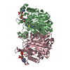
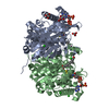
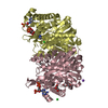
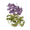
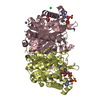
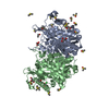
- PDB-4ubv: Structure of the 3-ketoacyl-CoA thiolase FadA5 from M. tuberculos... -
+ Open data

- Basic information
| Entry | Database: PDB / ID: 4ubv | |||||||||||||||||||||||||||
|---|---|---|---|---|---|---|---|---|---|---|---|---|---|---|---|---|---|---|---|---|---|---|---|---|---|---|---|---|
| Title | Structure of the 3-ketoacyl-CoA thiolase FadA5 from M. tuberculosis with an partially acetylated cysteine in complex with acetyl-CoA and CoA | |||||||||||||||||||||||||||
 Components Components | Acetyl-CoA acetyltransferase FadA5 | |||||||||||||||||||||||||||
 Keywords Keywords |  TRANSFERASE / partially acetylated degradative thiolase / acetyl-CoA / CoA TRANSFERASE / partially acetylated degradative thiolase / acetyl-CoA / CoA | |||||||||||||||||||||||||||
| Function / homology |  Function and homology informationacetyl-CoA C-acyltransferase / acetyl-CoA C-acyltransferase activity / phenylacetate catabolic process / cholesterol catabolic process / fatty acid beta-oxidation / identical protein binding Function and homology informationacetyl-CoA C-acyltransferase / acetyl-CoA C-acyltransferase activity / phenylacetate catabolic process / cholesterol catabolic process / fatty acid beta-oxidation / identical protein bindingSimilarity search - Function | |||||||||||||||||||||||||||
| Biological species |   Mycobacterium tuberculosis (bacteria) Mycobacterium tuberculosis (bacteria) | |||||||||||||||||||||||||||
| Method | X-RAY DIFFRACTION / SYNCHROTRON / MOLECULAR REPLACEMENT / Resolution: 1.95 Å | |||||||||||||||||||||||||||
 Authors Authors | Schaefer, C.M. / Kisker, C. | |||||||||||||||||||||||||||
| Funding support |  Germany, Germany,  United States, 8items United States, 8items
| |||||||||||||||||||||||||||
 Citation Citation | Journal: Structure / Year: 2015 Title: FadA5 a Thiolase from Mycobacterium tuberculosis: A Steroid-Binding Pocket Reveals the Potential for Drug Development against Tuberculosis. Authors: Schaefer, C.M. / Lu, R. / Nesbitt, N.M. / Schiebel, J. / Sampson, N.S. / Kisker, C. | |||||||||||||||||||||||||||
| History |
|
- Structure visualization
| Structure viewer | Molecule: MolmilJmol/JSmol |
|---|
- Downloads & links
-Download
| PDBx/mmCIF format | 4ubv.cif.gz | 319.9 KB | Display | PDBx/mmCIF format |
|---|---|---|---|---|
| PDB format | pdb4ubv.ent.gz | 258 KB | Display | PDB format |
| PDBx/mmJSON format | 4ubv.json.gz | Tree view | PDBx/mmJSON format | |
| Others |  Other downloads Other downloads |
-Validation report
| Arichive directory | https://data.pdbj.org/pub/pdb/validation_reports/ub/4ubvftp://data.pdbj.org/pub/pdb/validation_reports/ub/4ubv | HTTPS FTP |
|---|
-Related structure data
| Related structure data |  4ubtC  4ubuC  4ubwC 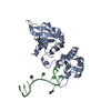 1u1qS S: Starting model for refinement C: citing same article ( |
|---|---|
| Similar structure data |


-Links
 PDBj
PDBj


- Assembly
| Deposited unit | 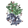
| ||||||||
|---|---|---|---|---|---|---|---|---|---|
| 1 |
| ||||||||
| Unit cell |
| ||||||||
| Components on special symmetry positions |
|

-Components
-Protein , 1 types, 2 molecules AB
| #1: Protein | Mass: 42346.723 Da / Num. of mol.: 2 Source method: isolated from a genetically manipulated source Details: THE THIOLASE FADA5 IN COMPLEX WITH ACETYL-COA AND COA; THE THIOLASE IS PARTIALLY COVALENTLY MODIFIED AT C93 (ACETYLATED) Source: (gene. exp.) Mycobacterium tuberculosis (bacteria) / Strain: ATCC 25618 / H37Rv / Gene: fadA5, Rv3546, P425_03688, RVBD_3546 / Plasmid: pSD31Production host: Mycobacterium smegmatis str. MC2 155 (bacteria)References: UniProt: I6XHI4 |
|---|
-Non-polymers , 6 types, 306 molecules 


| #2: Chemical | ChemComp-ACO / Acetyl-CoA Mass: 809.571 Da / Num. of mol.: 1 / Source method: obtained synthetically / Formula: C23H38N7O17P3S Mass: 809.571 Da / Num. of mol.: 1 / Source method: obtained synthetically / Formula: C23H38N7O17P3S | ||||||||
|---|---|---|---|---|---|---|---|---|---|
| #3: Chemical | Coenzyme A Mass: 767.534 Da / Num. of mol.: 2 / Source method: obtained synthetically / Formula: C21H36N7O16P3S Mass: 767.534 Da / Num. of mol.: 2 / Source method: obtained synthetically / Formula: C21H36N7O16P3S#4: Chemical | Sulfate Mass: 96.063 Da / Num. of mol.: 3 / Source method: obtained synthetically / Formula: SO4 Mass: 96.063 Da / Num. of mol.: 3 / Source method: obtained synthetically / Formula: SO4#5: Chemical | ChemComp-DIO / 1,4-Dioxane Mass: 88.105 Da / Num. of mol.: 7 / Source method: obtained synthetically / Formula: C4H8O2 Mass: 88.105 Da / Num. of mol.: 7 / Source method: obtained synthetically / Formula: C4H8O2#6: Chemical | ChemComp-GOL / Glycerol Mass: 92.094 Da / Num. of mol.: 12 / Source method: obtained synthetically / Formula: C3H8O3 Mass: 92.094 Da / Num. of mol.: 12 / Source method: obtained synthetically / Formula: C3H8O3#7: Water | ChemComp-HOH / | WaterMass: 18.015 Da / Num. of mol.: 281 / Source method: isolated from a natural source / Formula: H2O |
-Experimental details
-Experiment
| Experiment | Method: X-RAY DIFFRACTION |
|---|
- Sample preparation
| Crystal | Density Matthews: 2.86 Å3/Da / Density % sol: 57.01 % |
|---|---|
| Crystal grow | Temperature: 293 K / Method: vapor diffusion, sitting drop / pH: 6.5 / Details: 0.1 M Mes pH 6.5, 4% 1,4-dioxane, 1.8 M (NH4)2SO4 |
-Data collection
| Diffraction | Mean temperature: 100 K |
|---|---|
| Diffraction source | Source: SYNCHROTRON / Site: BESSY / Beamline: 14.1 / Wavelength: 0.9184 Å |
| Detector | Type: MARMOSAIC 225 mm CCD / Detector: CCD / Date: May 11, 2011 |
| Radiation | Protocol: SINGLE WAVELENGTH / Monochromatic (M) / Laue (L): M / Scattering type: x-ray |
| Radiation wavelength | Wavelength: 0.9184 Å / Relative weight: 1 |
| Reflection | Resolution: 1.95→35.99 Å / Num. obs: 72009 / % possible obs: 100 % / Redundancy: 14.6 % / Rmerge(I) obs: 0.133 / Net I/σ(I): 13.8 |
| Reflection shell | Resolution: 1.95→1.99 Å / Redundancy: 14.7 % / Mean I/σ(I) obs: 2.5 / % possible all: 100 |
- Processing
| Software |
| ||||||||||||||||||||||||||||||||||||||||||||||||||||||||||||||||||||||||||||||||||||||||||||||||||||||||||||||||||||||||||||||||||||||||||||||||||||||||||||||||||||||||||||||||||||||
|---|---|---|---|---|---|---|---|---|---|---|---|---|---|---|---|---|---|---|---|---|---|---|---|---|---|---|---|---|---|---|---|---|---|---|---|---|---|---|---|---|---|---|---|---|---|---|---|---|---|---|---|---|---|---|---|---|---|---|---|---|---|---|---|---|---|---|---|---|---|---|---|---|---|---|---|---|---|---|---|---|---|---|---|---|---|---|---|---|---|---|---|---|---|---|---|---|---|---|---|---|---|---|---|---|---|---|---|---|---|---|---|---|---|---|---|---|---|---|---|---|---|---|---|---|---|---|---|---|---|---|---|---|---|---|---|---|---|---|---|---|---|---|---|---|---|---|---|---|---|---|---|---|---|---|---|---|---|---|---|---|---|---|---|---|---|---|---|---|---|---|---|---|---|---|---|---|---|---|---|---|---|---|---|
| Refinement | Method to determine structure: MOLECULAR REPLACEMENT Starting model: 1u1q Resolution: 1.95→35.99 Å / Cor.coef. Fo:Fc: 0.935 / Cor.coef. Fo:Fc free: 0.915 / SU B: 6.58 / SU ML: 0.093 / Cross valid method: THROUGHOUT / ESU R: 0.15 / ESU R Free: 0.141 / Stereochemistry target values: MAXIMUM LIKELIHOOD / Details: HYDROGENS HAVE BEEN USED IF PRESENT IN THE INPUT
| ||||||||||||||||||||||||||||||||||||||||||||||||||||||||||||||||||||||||||||||||||||||||||||||||||||||||||||||||||||||||||||||||||||||||||||||||||||||||||||||||||||||||||||||||||||||
| Solvent computation | Solvent model: BABINET MODEL PARAMETERS FOR MASK CACLULATION | ||||||||||||||||||||||||||||||||||||||||||||||||||||||||||||||||||||||||||||||||||||||||||||||||||||||||||||||||||||||||||||||||||||||||||||||||||||||||||||||||||||||||||||||||||||||
| Displacement parameters | Biso mean: 29.824 Å2
| ||||||||||||||||||||||||||||||||||||||||||||||||||||||||||||||||||||||||||||||||||||||||||||||||||||||||||||||||||||||||||||||||||||||||||||||||||||||||||||||||||||||||||||||||||||||
| Refinement step | Cycle: 1 / Resolution: 1.95→35.99 Å
| ||||||||||||||||||||||||||||||||||||||||||||||||||||||||||||||||||||||||||||||||||||||||||||||||||||||||||||||||||||||||||||||||||||||||||||||||||||||||||||||||||||||||||||||||||||||
| Refine LS restraints |
|