 Movie
Movie Controller

[English] 日本語
 Yorodumi
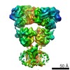
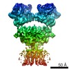
- PDB-4u4g: Structure of GluA2* in complex with competitive antagonist ZK 200775 -
+ Open data

- Basic information
| Entry | Database: PDB / ID: 4u4g | |||||||||
|---|---|---|---|---|---|---|---|---|---|---|
| Title | Structure of GluA2* in complex with competitive antagonist ZK 200775 | |||||||||
 Components Components | Glutamate receptor 2 GRIA2 GRIA2 | |||||||||
 Keywords Keywords | TRANSPORT PROTEIN / Ionotropic glutamate receptor / AMPA receptor / competitive antagonist / tetramer / complex | |||||||||
| Function / homology |  Function and homology information Function and homology informationspine synapse / dendritic spine neck / dendritic spine head / Activation of AMPA receptors / response to lithium ion / perisynaptic space / cellular response to glycine / AMPA glutamate receptor activity / Trafficking of GluR2-containing AMPA receptors / immunoglobulin binding ...spine synapse / dendritic spine neck / dendritic spine head / Activation of AMPA receptors / response to lithium ion / perisynaptic space / cellular response to glycine / AMPA glutamate receptor activity / Trafficking of GluR2-containing AMPA receptors / immunoglobulin binding / AMPA glutamate receptor complex / kainate selective glutamate receptor activity / ionotropic glutamate receptor complex / extracellularly glutamate-gated ion channel activity / asymmetric synapse / regulation of receptor recycling / Unblocking of NMDA receptors, glutamate binding and activation / glutamate receptor binding / positive regulation of synaptic transmission / presynaptic active zone membrane / glutamate-gated receptor activity / response to fungicide / regulation of synaptic transmission, glutamatergic / cellular response to brain-derived neurotrophic factor stimulus / somatodendritic compartment / dendrite membrane / ligand-gated monoatomic ion channel activity involved in regulation of presynaptic membrane potential / ionotropic glutamate receptor binding / cytoskeletal protein binding / ionotropic glutamate receptor signaling pathway / dendrite cytoplasm / SNARE binding / dendritic shaft / transmitter-gated monoatomic ion channel activity involved in regulation of postsynaptic membrane potential / synaptic membrane / synaptic transmission, glutamatergic / PDZ domain binding / postsynaptic density membrane / protein tetramerization / modulation of chemical synaptic transmission / Schaffer collateral - CA1 synapse / establishment of protein localization / terminal bouton / receptor internalization / synaptic vesicle membrane / cerebral cortex development / synaptic vesicle / presynapse / signaling receptor activity / presynaptic membrane / amyloid-beta binding / growth cone / chemical synaptic transmission / perikaryon / scaffold protein binding / postsynaptic membrane / dendritic spine / postsynaptic density / neuron projection / axon / dendrite / neuronal cell body / glutamatergic synapse / synapse / protein-containing complex binding / endoplasmic reticulum membrane / protein kinase binding / cell surface / endoplasmic reticulum / protein-containing complex / membrane / identical protein binding / plasma membraneSimilarity search - Function | |||||||||
| Biological species |  Rattus norvegicus (Norway rat) Rattus norvegicus (Norway rat) | |||||||||
| Method | X-RAY DIFFRACTION / SYNCHROTRON / MOLECULAR REPLACEMENT / Resolution: 4.49 Å | |||||||||
 Authors Authors | Yelshanskaya, M.V. / Li, M. / Sobolevsky, A.I. | |||||||||
| Funding support |  United States, 1items United States, 1items
| |||||||||
 Citation Citation | Journal: Science / Year: 2014 Title: Structure of an agonist-bound ionotropic glutamate receptor. Authors: Yelshanskaya, M.V. / Li, M. / Sobolevsky, A.I. | |||||||||
| History |
|
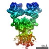
- Structure visualization
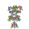
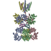
| Structure viewer | Molecule: MolmilJmol/JSmol |
|---|
- Downloads & links
-Download
| PDBx/mmCIF format | 4u4g.cif.gz | 532.9 KB | Display | PDBx/mmCIF format |
|---|---|---|---|---|
| PDB format | pdb4u4g.ent.gz | 439 KB | Display | PDB format |
| PDBx/mmJSON format | 4u4g.json.gz | Tree view | PDBx/mmJSON format | |
| Others |  Other downloads Other downloads |
-Validation report
| Arichive directory | https://data.pdbj.org/pub/pdb/validation_reports/u4/4u4gftp://data.pdbj.org/pub/pdb/validation_reports/u4/4u4g | HTTPS FTP |
|---|
-Related structure data
| Related structure data |  4u4fC 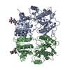 3h5vS 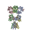 3kg2S C: citing same article ( S: Starting model for refinement |
|---|---|
| Similar structure data |


-Links
 PDBj
PDBj


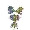
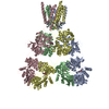
- Assembly
| Deposited unit | 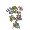
| ||||||||
|---|---|---|---|---|---|---|---|---|---|
| 1 |
| ||||||||
| Unit cell |
|
-Components
| #1: Protein | GRIA2 / GluR-2 / AMPA-selective glutamate receptor 2 / GluR-B / GluR-K2 / Glutamate receptor ionotropic / ...GluR-2 / AMPA-selective glutamate receptor 2 / GluR-B / GluR-K2 / Glutamate receptor ionotropic / AMPA 2 / GluA2 Mass: 92238.320 Da / Num. of mol.: 4 / Fragment: UNP residues 25-847 Source method: isolated from a genetically manipulated source Source: (gene. exp.) Rattus norvegicus (Norway rat) / Gene: Gria2, Glur2 / Production host:   Spodoptera frugiperda (fall armyworm) / References: UniProt: P19491 Spodoptera frugiperda (fall armyworm) / References: UniProt: P19491#2: Polysaccharide | beta-D-mannopyranose-(1-3)-beta-D-mannopyranose-(1-4)-2-acetamido-2-deoxy-beta-D-glucopyranose-(1-4) ...beta-D-mannopyranose-(1-3)-beta-D-mannopyranose-(1-4)-2-acetamido-2-deoxy-beta-D-glucopyranose-(1-4)-2-acetamido-2-deoxy-beta-D-glucopyranose / Mass: 748.682 Da / Num. of mol.: 4Source method: isolated from a genetically manipulated source #3: Chemical | ChemComp-ZK1 / {[ Fanapanel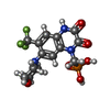  Mass: 409.254 Da / Num. of mol.: 4 / Source method: obtained synthetically / Formula: C14H15F3N3O6P / Comment: antagonist, medication*YM Mass: 409.254 Da / Num. of mol.: 4 / Source method: obtained synthetically / Formula: C14H15F3N3O6P / Comment: antagonist, medication*YM |
|---|
-Experimental details
-Experiment
| Experiment | Method: X-RAY DIFFRACTION / Number of used crystals: 1 |
|---|
- Sample preparation
| Crystal | Density Matthews: 4.17 Å3/Da / Density % sol: 70.51 % |
|---|---|
| Crystal grow | Temperature: 277 K / Method: batch mode / Details: 7-11% PEG 20,000, 0.1 M MES / PH range: 6.0-6.5 |
-Data collection
| Diffraction | Mean temperature: 100 K |
|---|---|
| Diffraction source | Source: SYNCHROTRON / Site: APS / Beamline: 24-ID-E / Wavelength: 0.9792 Å |
| Detector | Type: ADSC QUANTUM 315 / Detector: CCD / Date: Dec 17, 2012 |
| Radiation | Protocol: SINGLE WAVELENGTH / Monochromatic (M) / Laue (L): M / Scattering type: x-ray |
| Radiation wavelength | Wavelength: 0.9792 Å / Relative weight: 1 |
| Reflection | Resolution: 4.49→50 Å / Num. obs: 36039 / % possible obs: 99.8 % / Redundancy: 3.8 % / Biso Wilson estimate: 206.6 Å2 / Rmerge(I) obs: 0.098 / Net I/σ(I): 12.7 |
| Reflection shell | Resolution: 4.49→4.65 Å / Redundancy: 3.8 % / Rmerge(I) obs: 0.997 / Mean I/σ(I) obs: 1.48 / % possible all: 100 |
- Processing
| Software |
| ||||||||||||||||||||||||||||||||||||||||||||||||||||||||||||||||||||||||||||||||||||||||||||||||||
|---|---|---|---|---|---|---|---|---|---|---|---|---|---|---|---|---|---|---|---|---|---|---|---|---|---|---|---|---|---|---|---|---|---|---|---|---|---|---|---|---|---|---|---|---|---|---|---|---|---|---|---|---|---|---|---|---|---|---|---|---|---|---|---|---|---|---|---|---|---|---|---|---|---|---|---|---|---|---|---|---|---|---|---|---|---|---|---|---|---|---|---|---|---|---|---|---|---|---|---|
| Refinement | Method to determine structure: MOLECULAR REPLACEMENT Starting model: 3H5V, 3KG2 Resolution: 4.49→50 Å / SU ML: 0.59 / Cross valid method: FREE R-VALUE / σ(F): 1.36 / Phase error: 28.81 / Stereochemistry target values: ML
| ||||||||||||||||||||||||||||||||||||||||||||||||||||||||||||||||||||||||||||||||||||||||||||||||||
| Solvent computation | Shrinkage radii: 0.9 Å / VDW probe radii: 1.11 Å / Solvent model: FLAT BULK SOLVENT MODEL | ||||||||||||||||||||||||||||||||||||||||||||||||||||||||||||||||||||||||||||||||||||||||||||||||||
| Refinement step | Cycle: LAST / Resolution: 4.49→50 Å
| ||||||||||||||||||||||||||||||||||||||||||||||||||||||||||||||||||||||||||||||||||||||||||||||||||
| Refine LS restraints |
| ||||||||||||||||||||||||||||||||||||||||||||||||||||||||||||||||||||||||||||||||||||||||||||||||||
| LS refinement shell |
|
