 Movie
Movie Controller

[English] 日本語
 Yorodumi
- PDB-4tnl: 1.8 A resolution room temperature structure of Thermolysin record... -
+ Open data

- Basic information
| Entry | Database: PDB / ID: 4tnl | ||||||
|---|---|---|---|---|---|---|---|
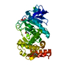
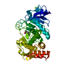
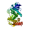
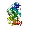
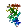
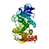
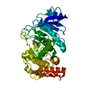
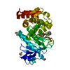
| Title | 1.8 A resolution room temperature structure of Thermolysin recorded using an XFEL | ||||||
 Components Components | Thermolysin | ||||||
 Keywords Keywords | HYDROLASE / Zn protease / X-ray free electron laser | ||||||
| Function / homology |  Function and homology informationthermolysin / metalloendopeptidase activity / proteolysis / extracellular region / metal ion binding Function and homology informationthermolysin / metalloendopeptidase activity / proteolysis / extracellular region / metal ion bindingSimilarity search - Function | ||||||
| Biological species |  | ||||||
| Method | X-RAY DIFFRACTION / FREE ELECTRON LASER / MOLECULAR REPLACEMENT / Resolution: 1.8 Å | ||||||
 Authors Authors | Kern, J. / Tran, R. / Alonso-Mori, R. / Koroidov, S. / Echols, N. / Hattne, J. / Ibrahim, M. / Gul, S. / Laksmono, H. / Sierra, R.G. ...Kern, J. / Tran, R. / Alonso-Mori, R. / Koroidov, S. / Echols, N. / Hattne, J. / Ibrahim, M. / Gul, S. / Laksmono, H. / Sierra, R.G. / Gildea, R.J. / Han, G. / Hellmich, J. / Lassalle-Kaiser, B. / Chatterjee, R. / Brewster, A. / Stan, C.A. / Gloeckner, C. / Lampe, A. / DiFiore, D. / Milathianaki, D. / Fry, A.R. / Seibert, M.M. / Koglin, J.E. / Gallo, E. / Uhlig, J. / Sokaras, D. / Weng, T.-C. / Zwart, P.H. / Skinner, D.E. / Bogan, M.J. / Messerschmidt, M. / Glatzel, P. / Williams, G.J. / Boutet, S. / Adams, P.D. / Zouni, A. / Messinger, J. / Sauter, N.K. / Bergmann, U. / Yano, J. / Yachandra, V.K. | ||||||
 Citation Citation | Journal: Nat Commun / Year: 2014 Title: Taking snapshots of photosynthetic water oxidation using femtosecond X-ray diffraction and spectroscopy. Authors: Kern, J. / Tran, R. / Alonso-Mori, R. / Koroidov, S. / Echols, N. / Hattne, J. / Ibrahim, M. / Gul, S. / Laksmono, H. / Sierra, R.G. / Gildea, R.J. / Han, G. / Hellmich, J. / Lassalle- ...Authors: Kern, J. / Tran, R. / Alonso-Mori, R. / Koroidov, S. / Echols, N. / Hattne, J. / Ibrahim, M. / Gul, S. / Laksmono, H. / Sierra, R.G. / Gildea, R.J. / Han, G. / Hellmich, J. / Lassalle-Kaiser, B. / Chatterjee, R. / Brewster, A.S. / Stan, C.A. / Glockner, C. / Lampe, A. / DiFiore, D. / Milathianaki, D. / Fry, A.R. / Seibert, M.M. / Koglin, J.E. / Gallo, E. / Uhlig, J. / Sokaras, D. / Weng, T.C. / Zwart, P.H. / Skinner, D.E. / Bogan, M.J. / Messerschmidt, M. / Glatzel, P. / Williams, G.J. / Boutet, S. / Adams, P.D. / Zouni, A. / Messinger, J. / Sauter, N.K. / Bergmann, U. / Yano, J. / Yachandra, V.K. #1: Journal: Acta Crystallogr. D Biol. Crystallogr. / Year: 2012 Title: Towards automated crystallographic structure refinement with phenix.refine. Authors: Afonine, P.V. / Grosse-Kunstleve, R.W. / Echols, N. / Headd, J.J. / Moriarty, N.W. / Mustyakimov, M. / Terwilliger, T.C. / Urzhumtsev, A. / Zwart, P.H. / Adams, P.D. #2: Journal: Acta Crystallogr D Biol Crystallogr / Year: 2010 Title: PHENIX: a comprehensive Python-based system for macromolecular structure solution. Authors: Paul D Adams / Pavel V Afonine / Gábor Bunkóczi / Vincent B Chen / Ian W Davis / Nathaniel Echols / Jeffrey J Headd / Li-Wei Hung / Gary J Kapral / Ralf W Grosse-Kunstleve / Airlie J McCoy ...Authors: Paul D Adams / Pavel V Afonine / Gábor Bunkóczi / Vincent B Chen / Ian W Davis / Nathaniel Echols / Jeffrey J Headd / Li-Wei Hung / Gary J Kapral / Ralf W Grosse-Kunstleve / Airlie J McCoy / Nigel W Moriarty / Robert Oeffner / Randy J Read / David C Richardson / Jane S Richardson / Thomas C Terwilliger / Peter H Zwart /  Abstract: Macromolecular X-ray crystallography is routinely applied to understand biological processes at a molecular level. However, significant time and effort are still required to solve and complete many ...Macromolecular X-ray crystallography is routinely applied to understand biological processes at a molecular level. However, significant time and effort are still required to solve and complete many of these structures because of the need for manual interpretation of complex numerical data using many software packages and the repeated use of interactive three-dimensional graphics. PHENIX has been developed to provide a comprehensive system for macromolecular crystallographic structure solution with an emphasis on the automation of all procedures. This has relied on the development of algorithms that minimize or eliminate subjective input, the development of algorithms that automate procedures that are traditionally performed by hand and, finally, the development of a framework that allows a tight integration between the algorithms. | ||||||
| History |
|
- Structure visualization
| Structure viewer | Molecule: MolmilJmol/JSmol |
|---|
- Downloads & links
-Download
| PDBx/mmCIF format | 4tnl.cif.gz | 141.6 KB | Display | PDBx/mmCIF format |
|---|---|---|---|---|
| PDB format | pdb4tnl.ent.gz | 103.1 KB | Display | PDB format |
| PDBx/mmJSON format | 4tnl.json.gz | Tree view | PDBx/mmJSON format | |
| Others |  Other downloads Other downloads |
-Validation report
| Arichive directory | https://data.pdbj.org/pub/pdb/validation_reports/tn/4tnlftp://data.pdbj.org/pub/pdb/validation_reports/tn/4tnl | HTTPS FTP |
|---|
-Related structure data
| Related structure data |  4tnhC  4tniC  4tnjC  4tnkC  2tliS S: Starting model for refinement C: citing same article ( |
|---|---|
| Similar structure data |


-Links
 PDBj
PDBj- Assembly
| Deposited unit | 
| ||||||||||||
|---|---|---|---|---|---|---|---|---|---|---|---|---|---|
| 1 |
| ||||||||||||
| Unit cell |
| ||||||||||||
| Components on special symmetry positions |
|
-Components
| #1: Protein | / Thermostable neutral proteinase Mass: 34360.336 Da / Num. of mol.: 1 / Fragment: UNP residues 233-548 / Source method: isolated from a natural source / Source: (natural) thermolysin | ||
|---|---|---|---|
| #2: Chemical | ChemComp-ZN /   Mass: 65.409 Da / Num. of mol.: 1 / Source method: obtained synthetically / Formula: Zn Mass: 65.409 Da / Num. of mol.: 1 / Source method: obtained synthetically / Formula: Zn | ||
| #3: Chemical | ChemComp-CA /   Mass: 40.078 Da / Num. of mol.: 4 / Source method: obtained synthetically / Formula: Ca Mass: 40.078 Da / Num. of mol.: 4 / Source method: obtained synthetically / Formula: Ca#4: Water | ChemComp-HOH / | Water Mass: 18.015 Da / Num. of mol.: 324 / Source method: isolated from a natural source / Formula: H2O Mass: 18.015 Da / Num. of mol.: 324 / Source method: isolated from a natural source / Formula: H2O |
-Experimental details
-Experiment
| Experiment | Method: X-RAY DIFFRACTION / Number of used crystals: 120408 |
|---|
- Sample preparation
| Crystal | Density Matthews: 2.39 Å3/Da / Density % sol: 48.47 % |
|---|---|
| Crystal grow | Temperature: 298 K / Method: batch mode Details: 300 ul of the protein stock was mixed in a 1:1 ratio with 40% PEG 2000, 100 mM MES pH 6.5, 5 mM CaCl2. Crystallization occurred within minutes. |
-Data collection
| Diffraction | Mean temperature: 298 K |
|---|---|
| Diffraction source | Source: FREE ELECTRON LASER / Site: SLAC LCLS / Beamline: CXI / Wavelength: 1.27 Å |
| Detector | Type: CS-PAD detector / Detector: PIXEL / Date: Mar 3, 2013 Details: Collected at the CXI instrument at LCLS/SLAC using the CSPAD |
| Radiation | Monochromator: No monochromator, FEL beam with 20-30 eV bandwidth Protocol: SINGLE WAVELENGTH / Monochromatic (M) / Laue (L): M / Scattering type: x-ray |
| Radiation wavelength | Wavelength: 1.27 Å / Relative weight: 1 |
| Reflection | Resolution: 1.8→34.27 Å / Num. obs: 31458 / % possible obs: 100 % / Redundancy: 1468 % / Biso Wilson estimate: 16.3967395539 Å2 / Net I/σ(I): 71.7 |
| Reflection shell | Resolution: 1.8→1.86 Å / Redundancy: 14.6 % / % possible all: 100 |
- Processing
| Software |
| |||||||||||||||||||||||||||||||||||||||||||||||||||||||||||||||||||||||||||||||||||||||||||||||||||||||||||||||||||||||||||||||||||||||||||||||||||||||||||||||||
|---|---|---|---|---|---|---|---|---|---|---|---|---|---|---|---|---|---|---|---|---|---|---|---|---|---|---|---|---|---|---|---|---|---|---|---|---|---|---|---|---|---|---|---|---|---|---|---|---|---|---|---|---|---|---|---|---|---|---|---|---|---|---|---|---|---|---|---|---|---|---|---|---|---|---|---|---|---|---|---|---|---|---|---|---|---|---|---|---|---|---|---|---|---|---|---|---|---|---|---|---|---|---|---|---|---|---|---|---|---|---|---|---|---|---|---|---|---|---|---|---|---|---|---|---|---|---|---|---|---|---|---|---|---|---|---|---|---|---|---|---|---|---|---|---|---|---|---|---|---|---|---|---|---|---|---|---|---|---|---|---|---|---|
| Refinement | Method to determine structure: MOLECULAR REPLACEMENT Starting model: 2tli Resolution: 1.8→34.27 Å / SU ML: 0.204817582335 / Cross valid method: FREE R-VALUE / σ(F): 1.42149651097 / Phase error: 20.1997292845
| |||||||||||||||||||||||||||||||||||||||||||||||||||||||||||||||||||||||||||||||||||||||||||||||||||||||||||||||||||||||||||||||||||||||||||||||||||||||||||||||||
| Solvent computation | Shrinkage radii: 0.9 Å / VDW probe radii: 1.11 Å / Solvent model: FLAT BULK SOLVENT MODEL | |||||||||||||||||||||||||||||||||||||||||||||||||||||||||||||||||||||||||||||||||||||||||||||||||||||||||||||||||||||||||||||||||||||||||||||||||||||||||||||||||
| Displacement parameters | Biso mean: 20.1244048785 Å2 | |||||||||||||||||||||||||||||||||||||||||||||||||||||||||||||||||||||||||||||||||||||||||||||||||||||||||||||||||||||||||||||||||||||||||||||||||||||||||||||||||
| Refinement step | Cycle: LAST / Resolution: 1.8→34.27 Å
| |||||||||||||||||||||||||||||||||||||||||||||||||||||||||||||||||||||||||||||||||||||||||||||||||||||||||||||||||||||||||||||||||||||||||||||||||||||||||||||||||
| Refine LS restraints |
| |||||||||||||||||||||||||||||||||||||||||||||||||||||||||||||||||||||||||||||||||||||||||||||||||||||||||||||||||||||||||||||||||||||||||||||||||||||||||||||||||
| LS refinement shell |
|
