 Movie
Movie Controller

[English] 日本語
 Yorodumi
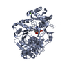
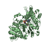
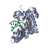
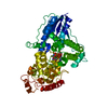
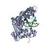
- PDB-4rv2: Crystal Structure of (3R)-hydroxyacyl-ACP dehydratase HadAB heter... -
+ Open data

- Basic information
| Entry | Database: PDB / ID: 4rv2 | ||||||
|---|---|---|---|---|---|---|---|
| Title | Crystal Structure of (3R)-hydroxyacyl-ACP dehydratase HadAB hetero-dimer from Mycobacterium smegmatis | ||||||
 Components Components |
| ||||||
 Keywords Keywords |  LYASE / Hotdog fold LYASE / Hotdog fold | ||||||
| Function / homology |  Function and homology informationDehydratase subunit HadA-like / N-terminal of MaoC-like dehydratase / N-terminal half of MaoC dehydratase / MaoC-like dehydratase domain / MaoC like domain / Hotdog Thioesterase / Thiol Ester Dehydrase; Chain A / HotDog domain superfamily / Roll / Alpha Beta Function and homology informationDehydratase subunit HadA-like / N-terminal of MaoC-like dehydratase / N-terminal half of MaoC dehydratase / MaoC-like dehydratase domain / MaoC like domain / Hotdog Thioesterase / Thiol Ester Dehydrase; Chain A / HotDog domain superfamily / Roll / Alpha BetaSimilarity search - Domain/homology | ||||||
| Biological species |  Mycobacterium smegmatis str. MC2 155 (bacteria) Mycobacterium smegmatis str. MC2 155 (bacteria) | ||||||
| Method | X-RAY DIFFRACTION / MIR / Resolution: 2.7 Å | ||||||
 Authors Authors | Biswas, R. / Hazra, D. / Dutta, D. / Das, A.K. | ||||||
 Citation Citation | Journal: Biochem.Biophys.Res.Commun. / Year: 2015 Title: Crystal structure of dehydratase component HadAB complex of mycobacterial FAS-II pathway. Authors: Biswas, R. / Dutta, A. / Dutta, D. / Hazra, D. / Banerjee, D.R. / Basak, A. / Das, A.K. | ||||||
| History |
|
- Structure visualization
| Structure viewer | Molecule: MolmilJmol/JSmol |
|---|
- Downloads & links
-Download
| PDBx/mmCIF format | 4rv2.cif.gz | 117.5 KB | Display | PDBx/mmCIF format |
|---|---|---|---|---|
| PDB format | pdb4rv2.ent.gz | 92.4 KB | Display | PDB format |
| PDBx/mmJSON format | 4rv2.json.gz | Tree view | PDBx/mmJSON format | |
| Others |  Other downloads Other downloads |
-Validation report
| Arichive directory | https://data.pdbj.org/pub/pdb/validation_reports/rv/4rv2ftp://data.pdbj.org/pub/pdb/validation_reports/rv/4rv2 | HTTPS FTP |
|---|
-Related structure data
| Similar structure data |
|---|


-Links
 PDBj
PDBj
- Assembly
| Deposited unit | 
| ||||||||
|---|---|---|---|---|---|---|---|---|---|
| 1 | 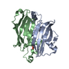
| ||||||||
| Unit cell |
|
-Components
| #1: Protein | Mass: 16622.625 Da / Num. of mol.: 1 / Fragment: UNP residues 7-144 Source method: isolated from a genetically manipulated source Source: (gene. exp.) Mycobacterium smegmatis str. MC2 155 (bacteria)Strain: mc2 155 / Gene: MSMEG_1340, MSMEI_1302 / Plasmid: pQE30 / Production host: Escherichia coli (E. coli) / Strain (production host): M15 / References: UniProt: A0QS40 |
|---|---|
| #2: Protein | Mass: 14741.694 Da / Num. of mol.: 1 / Fragment: UNP residues 2-142 Source method: isolated from a genetically manipulated source Source: (gene. exp.) Mycobacterium smegmatis str. MC2 155 (bacteria)Strain: mc2 155 / Gene: MSMEG_1341, MSMEI_1303 / Plasmid: pQE30 / Production host: Escherichia coli (E. coli) / Strain (production host): M15 / References: UniProt: A0QS41 |
| #3: Chemical | ChemComp-SO4 / Sulfate  Mass: 96.063 Da / Num. of mol.: 1 / Source method: obtained synthetically / Formula: SO4 Mass: 96.063 Da / Num. of mol.: 1 / Source method: obtained synthetically / Formula: SO4 |
| #4: Water | ChemComp-HOH / Water Mass: 18.015 Da / Num. of mol.: 53 / Source method: isolated from a natural source / Formula: H2O Mass: 18.015 Da / Num. of mol.: 53 / Source method: isolated from a natural source / Formula: H2O |
-Experimental details
-Experiment
| Experiment | Method: X-RAY DIFFRACTION / Number of used crystals: 1 |
|---|
- Sample preparation
| Crystal | Density Matthews: 3.08 Å3/Da / Density % sol: 60.08 % |
|---|---|
| Crystal grow | Temperature: 298 K / Method: vapor diffusion, hanging drop / pH: 6.8 Details: 1.75 M (NH4)2SO4, 0.1 M HEPES, 2% PEG 400, pH 6.8, VAPOR DIFFUSION, HANGING DROP, temperature 298K |
-Data collection
| Diffraction | Mean temperature: 100 K | ||||||||||||||||||||||||||||||||||||||||||||||||||||||||||||||||||||||||||||||||||||||||
|---|---|---|---|---|---|---|---|---|---|---|---|---|---|---|---|---|---|---|---|---|---|---|---|---|---|---|---|---|---|---|---|---|---|---|---|---|---|---|---|---|---|---|---|---|---|---|---|---|---|---|---|---|---|---|---|---|---|---|---|---|---|---|---|---|---|---|---|---|---|---|---|---|---|---|---|---|---|---|---|---|---|---|---|---|---|---|---|---|---|
| Diffraction source | Source: ROTATING ANODE / Type: RIGAKU MICROMAX-007 HF / Wavelength: 1.5418 Å | ||||||||||||||||||||||||||||||||||||||||||||||||||||||||||||||||||||||||||||||||||||||||
| Detector | Type: RIGAKU RAXIS IV++ / Detector: IMAGE PLATE / Date: Sep 12, 2014 / Details: Mirrors | ||||||||||||||||||||||||||||||||||||||||||||||||||||||||||||||||||||||||||||||||||||||||
| Radiation | Monochromator: Varimax, Osmic mirror / Protocol: SINGLE WAVELENGTH / Monochromatic (M) / Laue (L): M / Scattering type: x-ray | ||||||||||||||||||||||||||||||||||||||||||||||||||||||||||||||||||||||||||||||||||||||||
| Radiation wavelength | Wavelength: 1.5418 Å / Relative weight: 1 | ||||||||||||||||||||||||||||||||||||||||||||||||||||||||||||||||||||||||||||||||||||||||
| Reflection | Resolution: 2.699→19.732 Å / Num. all: 11408 / Num. obs: 11408 / % possible obs: 99.7 % / Redundancy: 41.8 % / Rsym value: 0.158 / Net I/σ(I): 36.2 | ||||||||||||||||||||||||||||||||||||||||||||||||||||||||||||||||||||||||||||||||||||||||
| Reflection shell | Diffraction-ID: 1
|
- Processing
| Software |
| |||||||||||||||||||||||||||||||||||||||||||||||||||||||||||||||||||||||||||
|---|---|---|---|---|---|---|---|---|---|---|---|---|---|---|---|---|---|---|---|---|---|---|---|---|---|---|---|---|---|---|---|---|---|---|---|---|---|---|---|---|---|---|---|---|---|---|---|---|---|---|---|---|---|---|---|---|---|---|---|---|---|---|---|---|---|---|---|---|---|---|---|---|---|---|---|---|
| Refinement | Method to determine structure: MIR / Resolution: 2.7→19.73 Å / Cor.coef. Fo:Fc: 0.922 / Cor.coef. Fo:Fc free: 0.86 / WRfactor Rfree: 0.2232 / WRfactor Rwork: 0.1739 / FOM work R set: 0.8054 / SU B: 23.032 / SU ML: 0.241 / SU R Cruickshank DPI: 0.5401 / SU Rfree: 0.3432 / Cross valid method: THROUGHOUT / σ(F): 0 / ESU R: 0.54 / ESU R Free: 0.343 / Stereochemistry target values: MAXIMUM LIKELIHOOD Details: HYDROGENS HAVE BEEN ADDED IN THE RIDING POSITIONS U VALUES
| |||||||||||||||||||||||||||||||||||||||||||||||||||||||||||||||||||||||||||
| Solvent computation | Ion probe radii: 0.8 Å / Shrinkage radii: 0.8 Å / VDW probe radii: 1.2 Å / Solvent model: MASK | |||||||||||||||||||||||||||||||||||||||||||||||||||||||||||||||||||||||||||
| Displacement parameters | Biso max: 57.15 Å2 / Biso mean: 34.629 Å2 / Biso min: 6.3 Å2
| |||||||||||||||||||||||||||||||||||||||||||||||||||||||||||||||||||||||||||
| Refinement step | Cycle: LAST / Resolution: 2.7→19.73 Å
| |||||||||||||||||||||||||||||||||||||||||||||||||||||||||||||||||||||||||||
| Refine LS restraints |
| |||||||||||||||||||||||||||||||||||||||||||||||||||||||||||||||||||||||||||
| LS refinement shell | Resolution: 2.7→2.768 Å / Total num. of bins used: 20
| |||||||||||||||||||||||||||||||||||||||||||||||||||||||||||||||||||||||||||
| Refinement TLS params. | Method: refined / Refine-ID: X-RAY DIFFRACTION
| |||||||||||||||||||||||||||||||||||||||||||||||||||||||||||||||||||||||||||
| Refinement TLS group |
|
