 Movie
Movie Controller

[English] 日本語
 Yorodumi
- PDB-4rp3: Crystal Structure of the L27 Domain of Discs Large 1 (target ID N... -
+ Open data

- Basic information
| Entry | Database: PDB / ID: 4rp3 | ||||||
|---|---|---|---|---|---|---|---|
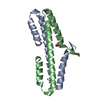
| Title | Crystal Structure of the L27 Domain of Discs Large 1 (target ID NYSGRC-010766) from Drosophila melanogaster bound to a potassium ion (space group P212121) | ||||||
 Components Components | Disks large 1 tumor suppressor protein | ||||||
 Keywords Keywords |  ANTITUMOR PROTEIN / NYSGRC / Structural Genomics / PSI-Biology / New York Structural Genomics Research Consortium / Scaffolding ANTITUMOR PROTEIN / NYSGRC / Structural Genomics / PSI-Biology / New York Structural Genomics Research Consortium / Scaffolding | ||||||
| Function / homology |  Function and homology information Function and homology informationsmooth septate junction / positive phototaxis / RHO GTPases activate CIT / Neurexins and neuroligins / establishment or maintenance of polarity of larval imaginal disc epithelium / cell fate commitment involved in pattern specification / type I terminal bouton / septate junction / septate junction assembly / negative regulation of imaginal disc growth ...smooth septate junction / positive phototaxis / RHO GTPases activate CIT / Neurexins and neuroligins / establishment or maintenance of polarity of larval imaginal disc epithelium / cell fate commitment involved in pattern specification / type I terminal bouton / septate junction / septate junction assembly / negative regulation of imaginal disc growth / Activation of Ca-permeable Kainate Receptor / morphogenesis of follicular epithelium / type Ib terminal bouton / subsynaptic reticulum / Synaptic adhesion-like molecules / establishment or maintenance of polarity of follicular epithelium / morphogenesis of larval imaginal disc epithelium / Unblocking of NMDA receptors, glutamate binding and activation / gravitaxis / tricellular tight junction / establishment of spindle orientation / anterior/posterior axis specification, follicular epithelium / negative regulation of peptidoglycan recognition protein signaling pathway / pole plasm protein localization / basal protein localization / asymmetric protein localization involved in cell fate determination / dorsal closure / follicle cell of egg chamber development / regulation of epidermal growth factor receptor signaling pathway / positive regulation of synaptic assembly at neuromuscular junction / male courtship behavior / guanylate kinase activity / receptor localization to synapse / morphogenesis of a polarized epithelium / behavioral response to ethanol / apical cortex / apicolateral plasma membrane / leading edge membrane / establishment or maintenance of epithelial cell apical/basal polarity / synaptic assembly at neuromuscular junction / mating behavior / cell fate specification / receptor clustering / locomotor rhythm / establishment of mitotic spindle orientation / lateral plasma membrane / postsynaptic density membrane / neuromuscular junction / protein localization / terminal bouton / cell-cell adhesion / kinase binding / nervous system development / cell cortex / chemical synaptic transmission / postsynaptic membrane / basolateral plasma membrane / cytoskeleton / neuron projection / synapse / perinuclear region of cytoplasm / signal transduction / plasma membraneSimilarity search - Function | ||||||
| Biological species |  Drosophila melanogaster (fruit fly) Drosophila melanogaster (fruit fly) | ||||||
| Method | X-RAY DIFFRACTION / SYNCHROTRON / SAD / Resolution: 1.36 Å | ||||||
 Authors Authors | Ghosh, A. / Ramagopal, U. / Almo, S.C. / New York Structural Genomics Research Consortium (NYSGRC) | ||||||
 Citation Citation | Journal: Biochemistry / Year: 2018 Title: Structures of the L27 Domain of Disc Large Homologue 1 Protein Illustrate a Self-Assembly Module. Authors: Ghosh, A. / Ramagopal, U.A. / Bonanno, J.B. / Brenowitz, M. / Almo, S.C. | ||||||
| History |
|
- Structure visualization
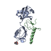
| Structure viewer | Molecule: MolmilJmol/JSmol |
|---|
- Downloads & links
-Download
| PDBx/mmCIF format | 4rp3.cif.gz | 98.6 KB | Display | PDBx/mmCIF format |
|---|---|---|---|---|
| PDB format | pdb4rp3.ent.gz | 75.6 KB | Display | PDB format |
| PDBx/mmJSON format | 4rp3.json.gz | Tree view | PDBx/mmJSON format | |
| Others |  Other downloads Other downloads |
-Validation report
| Arichive directory | https://data.pdbj.org/pub/pdb/validation_reports/rp/4rp3ftp://data.pdbj.org/pub/pdb/validation_reports/rp/4rp3 | HTTPS FTP |
|---|
-Related structure data


-Links
 PDBj
PDBj

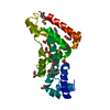
- Assembly
| Deposited unit | 
| ||||||||
|---|---|---|---|---|---|---|---|---|---|
| 1 |
| ||||||||
| Unit cell |
|
-Components
| #1: Protein | Mass: 11432.120 Da / Num. of mol.: 2 / Fragment: L27 domain residues 1-97 Source method: isolated from a genetically manipulated source Source: (gene. exp.) Drosophila melanogaster (fruit fly) / Gene: CG1725, Discs Large 1 Isoform B, dlg1, l(1)dlg1 / Plasmid: pSGC-His / Production host:  Escherichia coli BL21(DE3) (bacteria) / Strain (production host): BL21 (DE3) pRIL / References: UniProt: P31007 Escherichia coli BL21(DE3) (bacteria) / Strain (production host): BL21 (DE3) pRIL / References: UniProt: P31007#2: Chemical | ChemComp-K / |   Mass: 39.098 Da / Num. of mol.: 1 / Source method: obtained synthetically / Formula: K Mass: 39.098 Da / Num. of mol.: 1 / Source method: obtained synthetically / Formula: K#3: Chemical | Formic acid  Mass: 46.025 Da / Num. of mol.: 2 / Source method: obtained synthetically / Formula: CH2O2 Mass: 46.025 Da / Num. of mol.: 2 / Source method: obtained synthetically / Formula: CH2O2#4: Chemical | ChemComp-CL / | Chloride  Mass: 35.453 Da / Num. of mol.: 1 / Source method: obtained synthetically / Formula: Cl Mass: 35.453 Da / Num. of mol.: 1 / Source method: obtained synthetically / Formula: Cl#5: Water | ChemComp-HOH / | Water Mass: 18.015 Da / Num. of mol.: 163 / Source method: isolated from a natural source / Formula: H2O Mass: 18.015 Da / Num. of mol.: 163 / Source method: isolated from a natural source / Formula: H2O |
|---|
-Experimental details
-Experiment
| Experiment | Method: X-RAY DIFFRACTION / Number of used crystals: 1 |
|---|
- Sample preparation
| Crystal | Density Matthews: 1.95 Å3/Da / Density % sol: 36.89 % |
|---|---|
| Crystal grow | Temperature: 293 K / Method: vapor diffusion, sitting drop Details: 0.2 M Sodium Format, 20 % PEG 3350, VAPOR DIFFUSION, SITTING DROP, temperature 293K |
-Data collection
| Diffraction | Mean temperature: 77 K | |||||||||||||||||||||||||||||||||||||||||||||||||||||||||||||||||||||||||||||||||||||||||||||||||||||||||||||||||||||||||||||||||||||||||||||||||||
|---|---|---|---|---|---|---|---|---|---|---|---|---|---|---|---|---|---|---|---|---|---|---|---|---|---|---|---|---|---|---|---|---|---|---|---|---|---|---|---|---|---|---|---|---|---|---|---|---|---|---|---|---|---|---|---|---|---|---|---|---|---|---|---|---|---|---|---|---|---|---|---|---|---|---|---|---|---|---|---|---|---|---|---|---|---|---|---|---|---|---|---|---|---|---|---|---|---|---|---|---|---|---|---|---|---|---|---|---|---|---|---|---|---|---|---|---|---|---|---|---|---|---|---|---|---|---|---|---|---|---|---|---|---|---|---|---|---|---|---|---|---|---|---|---|---|---|---|---|
| Diffraction source | Source: SYNCHROTRON / Site: NSLS  / Beamline: X29A / Wavelength: 0.9786 Å / Beamline: X29A / Wavelength: 0.9786 Å | |||||||||||||||||||||||||||||||||||||||||||||||||||||||||||||||||||||||||||||||||||||||||||||||||||||||||||||||||||||||||||||||||||||||||||||||||||
| Detector | Type: ADSC QUANTUM 315 / Detector: CCD / Date: Jul 25, 2013 | |||||||||||||||||||||||||||||||||||||||||||||||||||||||||||||||||||||||||||||||||||||||||||||||||||||||||||||||||||||||||||||||||||||||||||||||||||
| Radiation | Monochromator: ROSENBAUM-ROCK DOUBLE CRYSTAL / Protocol: SINGLE WAVELENGTH / Monochromatic (M) / Laue (L): M / Scattering type: x-ray | |||||||||||||||||||||||||||||||||||||||||||||||||||||||||||||||||||||||||||||||||||||||||||||||||||||||||||||||||||||||||||||||||||||||||||||||||||
| Radiation wavelength | Wavelength: 0.9786 Å / Relative weight: 1 | |||||||||||||||||||||||||||||||||||||||||||||||||||||||||||||||||||||||||||||||||||||||||||||||||||||||||||||||||||||||||||||||||||||||||||||||||||
| Reflection | Redundancy: 7.2 % / Number: 280814 / Rmerge(I) obs: 0.059 / Χ2: 1.11 / D res high: 1.36 Å / D res low: 30 Å / Num. obs: 39086 / % possible obs: 99.9 | |||||||||||||||||||||||||||||||||||||||||||||||||||||||||||||||||||||||||||||||||||||||||||||||||||||||||||||||||||||||||||||||||||||||||||||||||||
| Diffraction reflection shell |
| |||||||||||||||||||||||||||||||||||||||||||||||||||||||||||||||||||||||||||||||||||||||||||||||||||||||||||||||||||||||||||||||||||||||||||||||||||
| Reflection | Resolution: 1.36→30 Å / Num. all: 39126 / Num. obs: 39086 / % possible obs: 99.9 % / Observed criterion σ(F): 0 / Observed criterion σ(I): 0 / Redundancy: 7.2 % / Rmerge(I) obs: 0.059 / Χ2: 1.11 / Net I/σ(I): 14.6 | |||||||||||||||||||||||||||||||||||||||||||||||||||||||||||||||||||||||||||||||||||||||||||||||||||||||||||||||||||||||||||||||||||||||||||||||||||
| Reflection shell |
|
- Processing
| Software |
| ||||||||||||||||||||||||||||||||||||||||||||||||||||||||||||||||||||||||||||||||||||||||||
|---|---|---|---|---|---|---|---|---|---|---|---|---|---|---|---|---|---|---|---|---|---|---|---|---|---|---|---|---|---|---|---|---|---|---|---|---|---|---|---|---|---|---|---|---|---|---|---|---|---|---|---|---|---|---|---|---|---|---|---|---|---|---|---|---|---|---|---|---|---|---|---|---|---|---|---|---|---|---|---|---|---|---|---|---|---|---|---|---|---|---|---|
| Refinement | Method to determine structure: SAD / Resolution: 1.36→30 Å / Cor.coef. Fo:Fc: 0.976 / Cor.coef. Fo:Fc free: 0.958 / SU B: 2.603 / SU ML: 0.046 / Cross valid method: THROUGHOUT / σ(F): 0 / ESU R: 0.057 / ESU R Free: 0.061 / Stereochemistry target values: MAXIMUM LIKELIHOOD Details: HYDROGENS HAVE BEEN ADDED IN THE RIDING POSITIONS U VALUES : REFINED INDIVIDUALLY
| ||||||||||||||||||||||||||||||||||||||||||||||||||||||||||||||||||||||||||||||||||||||||||
| Solvent computation | Ion probe radii: 0.8 Å / Shrinkage radii: 0.8 Å / VDW probe radii: 1.2 Å / Solvent model: MASK | ||||||||||||||||||||||||||||||||||||||||||||||||||||||||||||||||||||||||||||||||||||||||||
| Displacement parameters | Biso max: 183.07 Å2 / Biso mean: 27.565 Å2 / Biso min: 11.65 Å2
| ||||||||||||||||||||||||||||||||||||||||||||||||||||||||||||||||||||||||||||||||||||||||||
| Refinement step | Cycle: LAST / Resolution: 1.36→30 Å
| ||||||||||||||||||||||||||||||||||||||||||||||||||||||||||||||||||||||||||||||||||||||||||
| Refine LS restraints |
| ||||||||||||||||||||||||||||||||||||||||||||||||||||||||||||||||||||||||||||||||||||||||||
| LS refinement shell | Resolution: 1.36→1.394 Å / Total num. of bins used: 20
| ||||||||||||||||||||||||||||||||||||||||||||||||||||||||||||||||||||||||||||||||||||||||||
| Refinement TLS params. | L11: 0 °2 / L12: 0 °2 / L13: 0 °2 / L22: 0 °2 / L23: 0 °2 / L33: 0 °2 / S11: 0 Å ° / S12: 0 Å ° / S13: 0 Å ° / S21: 0 Å ° / S22: 0 Å ° / S23: 0 Å ° / S31: 0 Å ° / S32: 0 Å ° / S33: 0 Å ° / T11: 0 Å2 / T12: 0 Å2 / T13: 0 Å2 / T22: 0 Å2 / T23: 0 Å2 / T33: 0 Å2 / Method: refined / Refine-ID: X-RAY DIFFRACTION
| ||||||||||||||||||||||||||||||||||||||||||||||||||||||||||||||||||||||||||||||||||||||||||
| Refinement TLS group |
|
