- PDB-4rhw: Crystal structure of Apaf-1 CARD and caspase-9 CARD complex -
+
Open data
ID or keywords:
Loading...
-
Basic information
Entry
Database: PDB / ID: 4rhw
Title
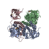
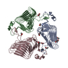
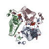
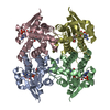
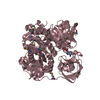
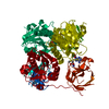
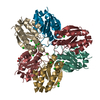
Crystal structure of Apaf-1 CARD and caspase-9 CARD complex
Components
Apoptotic protease-activating factor 1
Caspase-9
Keywords
APOPTOSIS / death domain superfamily
Function / homology
Function and homology information
caspase-9 / response to G1 DNA damage checkpoint signaling / caspase complex / regulation of apoptotic DNA fragmentation / Formation of apoptosome / apoptosome / activation of cysteine-type endopeptidase activity involved in apoptotic process by cytochrome c / leukocyte apoptotic process / glial cell apoptotic process / response to cobalt ion ...caspase-9 / response to G1 DNA damage checkpoint signaling / caspase complex / regulation of apoptotic DNA fragmentation / Formation of apoptosome / apoptosome / activation of cysteine-type endopeptidase activity involved in apoptotic process by cytochrome c / leukocyte apoptotic process / glial cell apoptotic process / response to cobalt ion / cysteine-type endopeptidase activity involved in apoptotic signaling pathway / Caspase activation via Dependence Receptors in the absence of ligand / Regulation of the apoptosome activity / Activation of caspases through apoptosome-mediated cleavage / cysteine-type endopeptidase activity involved in apoptotic process / SMAC (DIABLO) binds to IAPs / SMAC(DIABLO)-mediated dissociation of IAP:caspase complexes / epithelial cell apoptotic process / fibroblast apoptotic process / AKT phosphorylates targets in the cytosol / platelet formation / TP53 Regulates Transcription of Caspase Activators and Caspases / Transcriptional Regulation by E2F6 / Constitutive Signaling by AKT1 E17K in Cancer / cysteine-type endopeptidase activator activity involved in apoptotic process / protein maturation / enzyme activator activity / intrinsic apoptotic signaling pathway in response to endoplasmic reticulum stress / signal transduction in response to DNA damage / forebrain development / positive regulation of apoptotic signaling pathway / cardiac muscle cell apoptotic process / cellular response to transforming growth factor beta stimulus / heat shock protein binding / cellular response to dexamethasone stimulus / intrinsic apoptotic signaling pathway / response to nutrient / kidney development / neural tube closure / response to ischemia / ADP binding / NOD1/2 Signaling Pathway / protein processing / SH3 domain binding / activation of cysteine-type endopeptidase activity involved in apoptotic process / positive regulation of neuron apoptotic process / cellular response to UV / intrinsic apoptotic signaling pathway in response to DNA damage / response to estradiol / nervous system development / peptidase activity / neuron apoptotic process / regulation of apoptotic process / secretory granule lumen / ficolin-1-rich granule lumen / response to lipopolysaccharide / cell differentiation / response to hypoxia / positive regulation of apoptotic process / cysteine-type endopeptidase activity / nucleotide binding / apoptotic process / DNA damage response / Neutrophil degranulation / protein kinase binding / protein-containing complex / mitochondrion / extracellular exosome / extracellular region / ATP binding / identical protein binding / nucleus / cytosol / cytoplasm Similarity search - Function
AUTHOR STATED THE BIOLOGICAL UNIT CONTAINS 7 APAF-1 CARDS AND 4 CASPASE-9 CARDS. BASED ON THEIR EXPERIMENTAL RESULTS, APAF-1 FORMS A HEPTAMER IN THE APOPTOSOME, AND IN THE HOLOENZYME FORMED BY THE APOPTOSOME AND CASPASE-9, THE MOLAR RATIO BETWEEN APAF-1 AND CASPASE-9 IS 7:4. HOWEVER, FURTHER VALIDATION IS REQUIRED.
-
Components
#1: Protein
Apoptoticprotease-activatingfactor1 / APAF-1
Mass: 11099.710 Da / Num. of mol.: 4 / Fragment: card domain Source method: isolated from a genetically manipulated source Source: (gene. exp.) Homo sapiens (human) / Gene: APAF1, KIAA0413 / Production host: Escherichia coli (E. coli) / References: UniProt: O14727
In the structure databanks used in Yorodumi, some data are registered as the other names, "COVID-19 virus" and "2019-nCoV". Here are the details of the virus and the list of structure data.
Jan 31, 2019. EMDB accession codes are about to change! (news from PDBe EMDB page)
EMDB accession codes are about to change! (news from PDBe EMDB page)
The allocation of 4 digits for EMDB accession codes will soon come to an end. Whilst these codes will remain in use, new EMDB accession codes will include an additional digit and will expand incrementally as the available range of codes is exhausted. The current 4-digit format prefixed with “EMD-” (i.e. EMD-XXXX) will advance to a 5-digit format (i.e. EMD-XXXXX), and so on. It is currently estimated that the 4-digit codes will be depleted around Spring 2019, at which point the 5-digit format will come into force.
The EM Navigator/Yorodumi systems omit the EMD- prefix.
Related info.:Q: What is EMD? / ID/Accession-code notation in Yorodumi/EM Navigator
Yorodumi is a browser for structure data from EMDB, PDB, SASBDB, etc.
This page is also the successor to EM Navigator detail page, and also detail information page/front-end page for Omokage search.
The word "yorodu" (or yorozu) is an old Japanese word meaning "ten thousand". "mi" (miru) is to see.
Related info.:EMDB / PDB / SASBDB / Comparison of 3 databanks / Yorodumi Search / Aug 31, 2016. New EM Navigator & Yorodumi / Yorodumi Papers / Jmol/JSmol / Function and homology information / Changes in new EM Navigator and Yorodumi
 Movie
Movie Controller


 Open data
 Basic information
 Components
 Keywords
 APOPTOSIS /
  Function and homology information
 Authors
 Citation
 Structure visualization
 Downloads & links
 Other downloads
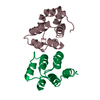

 PDBj
PDBj


 Assembly


 Mass: 96.063 Da / Num. of mol.: 7 / Source method: obtained synthetically / Formula: SO4
 Mass: 35.453 Da / Num. of mol.: 2 / Source method: obtained synthetically / Formula: Cl
 Mass: 18.015 Da / Num. of mol.: 355 / Source method: isolated from a natural source / Formula: H2O
 Sample preparation
 Processing