 Movie
Movie Controller

[English] 日本語
 Yorodumi
- PDB-4rb4: Crystal structure of dodecameric iron-containing heptosyltransfer... -
+ Open data

- Basic information
| Entry | Database: PDB / ID: 4rb4 | ||||||
|---|---|---|---|---|---|---|---|
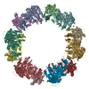
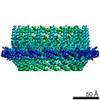
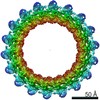
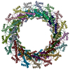
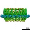
| Title | Crystal structure of dodecameric iron-containing heptosyltransferase TibC in complex with ADP-D-beta-D-heptose at 3.9 angstrom resolution | ||||||
 Components Components | Glycosyltransferase tibC | ||||||
 Keywords Keywords |  TRANSFERASE / GT-B fold / Heptose transfer / TibA / ADP-D-beta-D-heptose TRANSFERASE / GT-B fold / Heptose transfer / TibA / ADP-D-beta-D-heptose | ||||||
| Function / homology | Chem-AQH / : / :  Function and homology information Function and homology information | ||||||
| Biological species |  Escherichia coli DEC13E (bacteria) Escherichia coli DEC13E (bacteria) | ||||||
| Method | X-RAY DIFFRACTION / SYNCHROTRON / MOLECULAR REPLACEMENT / Resolution: 3.88 Å | ||||||
 Authors Authors | Yao, Q. / Lu, Q. / Shao, F. | ||||||
 Citation Citation | Journal: Elife / Year: 2014 Title: A structural mechanism for bacterial autotransporter glycosylation by a dodecameric heptosyltransferase family. Authors: Qing Yao / Qiuhe Lu / Xiaobo Wan / Feng Song / Yue Xu / Mo Hu / Alla Zamyatina / Xiaoyun Liu / Niu Huang / Ping Zhu / Feng Shao /   Abstract: A large group of bacterial virulence autotransporters including AIDA-I from diffusely adhering E. coli (DAEC) and TibA from enterotoxigenic E. coli (ETEC) require hyperglycosylation for functioning. ...A large group of bacterial virulence autotransporters including AIDA-I from diffusely adhering E. coli (DAEC) and TibA from enterotoxigenic E. coli (ETEC) require hyperglycosylation for functioning. Here we demonstrate that TibC from ETEC harbors a heptosyltransferase activity on TibA and AIDA-I, defining a large family of bacterial autotransporter heptosyltransferases (BAHTs). The crystal structure of TibC reveals a characteristic ring-shape dodecamer. The protomer features an N-terminal β-barrel, a catalytic domain, a β-hairpin thumb, and a unique iron-finger motif. The iron-finger motif contributes to back-to-back dimerization; six dimers form the ring through β-hairpin thumb-mediated hand-in-hand contact. The structure of ADP-D-glycero-β-D-manno-heptose (ADP-D,D-heptose)-bound TibC reveals a sugar transfer mechanism and also the ligand stereoselectivity determinant. Electron-cryomicroscopy analyses uncover a TibC-TibA dodecamer/hexamer assembly with two enzyme molecules binding to one TibA substrate. The complex structure also highlights a high efficient hyperglycosylation of six autotransporter substrates simultaneously by the dodecamer enzyme complex. | ||||||
| History |
|
- Structure visualization
| Structure viewer | Molecule: MolmilJmol/JSmol |
|---|
- Downloads & links
-Download
| PDBx/mmCIF format | 4rb4.cif.gz | 798.4 KB | Display | PDBx/mmCIF format |
|---|---|---|---|---|
| PDB format | pdb4rb4.ent.gz | 644.7 KB | Display | PDB format |
| PDBx/mmJSON format | 4rb4.json.gz | Tree view | PDBx/mmJSON format | |
| Others |  Other downloads Other downloads |
-Validation report
| Arichive directory | https://data.pdbj.org/pub/pdb/validation_reports/rb/4rb4ftp://data.pdbj.org/pub/pdb/validation_reports/rb/4rb4 | HTTPS FTP |
|---|
-Related structure data
| Related structure data | 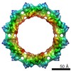 2755C 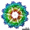 2756C  2757C 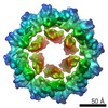 2758C  4rapSC S: Starting model for refinement C: citing same article ( |
|---|---|
| Similar structure data |


-Links
 PDBj
PDBj- Assembly
| Deposited unit | 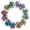
| ||||||||||||||||||||||||||||||||||||||||||||||||||||||||||||||||||||||||||||||||||||||||||||||||||||||||||||||||||||||||||||||||||||||||||||||
|---|---|---|---|---|---|---|---|---|---|---|---|---|---|---|---|---|---|---|---|---|---|---|---|---|---|---|---|---|---|---|---|---|---|---|---|---|---|---|---|---|---|---|---|---|---|---|---|---|---|---|---|---|---|---|---|---|---|---|---|---|---|---|---|---|---|---|---|---|---|---|---|---|---|---|---|---|---|---|---|---|---|---|---|---|---|---|---|---|---|---|---|---|---|---|---|---|---|---|---|---|---|---|---|---|---|---|---|---|---|---|---|---|---|---|---|---|---|---|---|---|---|---|---|---|---|---|---|---|---|---|---|---|---|---|---|---|---|---|---|---|---|---|---|
| 1 |
| ||||||||||||||||||||||||||||||||||||||||||||||||||||||||||||||||||||||||||||||||||||||||||||||||||||||||||||||||||||||||||||||||||||||||||||||
| Unit cell |
| ||||||||||||||||||||||||||||||||||||||||||||||||||||||||||||||||||||||||||||||||||||||||||||||||||||||||||||||||||||||||||||||||||||||||||||||
| Noncrystallographic symmetry (NCS) | NCS domain:
NCS domain segments: Component-ID: 1 / Ens-ID: 1 / End auth comp-ID: THR / End label comp-ID: THR / Refine code: 4
NCS oper:
|
-Components
| #1: Protein | Mass: 46334.500 Da / Num. of mol.: 12 / Mutation: D110A Source method: isolated from a genetically manipulated source Source: (gene. exp.) Escherichia coli DEC13E (bacteria) / Gene: tibC, ECDEC13E_2355 / Production host: Escherichia coli (E. coli)References: UniProt: H5MH13, Transferases; Glycosyltransferases#2: Chemical | ChemComp-FE / Iron  Mass: 55.845 Da / Num. of mol.: 12 / Source method: obtained synthetically / Formula: Fe Mass: 55.845 Da / Num. of mol.: 12 / Source method: obtained synthetically / Formula: Fe#3: Chemical | ChemComp-AQH / [(   Mass: 619.368 Da / Num. of mol.: 12 / Source method: obtained synthetically / Formula: C17H27N5O16P2 Mass: 619.368 Da / Num. of mol.: 12 / Source method: obtained synthetically / Formula: C17H27N5O16P2#4: Chemical | Ethylene glycol  Mass: 62.068 Da / Num. of mol.: 3 / Source method: obtained synthetically / Formula: C2H6O2 Mass: 62.068 Da / Num. of mol.: 3 / Source method: obtained synthetically / Formula: C2H6O2 |
|---|
-Experimental details
-Experiment
| Experiment | Method: X-RAY DIFFRACTION / Number of used crystals: 1 |
|---|
- Sample preparation
| Crystal | Density Matthews: 4.02 Å3/Da / Density % sol: 69.38 % |
|---|---|
| Crystal grow | Temperature: 298 K / Method: vapor diffusion, hanging drop / pH: 5.5 Details: 8% PEG 8000, 120mM magnesium acetate, 100mM MES buffer pH 5.5, VAPOR DIFFUSION, HANGING DROP, temperature 298K |
-Data collection
| Diffraction | Mean temperature: 200 K |
|---|---|
| Diffraction source | Source: SYNCHROTRON / Site: SSRF / Beamline: BL17U / Wavelength: 0.9791 Å |
| Detector | Type: ADSC QUANTUM 315r / Detector: CCD / Date: Jun 29, 2014 |
| Radiation | Monochromator: GRAPHITE / Protocol: SINGLE WAVELENGTH / Monochromatic (M) / Laue (L): M / Scattering type: x-ray |
| Radiation wavelength | Wavelength: 0.9791 Å / Relative weight: 1 |
| Reflection | Resolution: 3.88→20.04 Å / Num. all: 76251 / Num. obs: 75852 / % possible obs: 98.7 % / Observed criterion σ(F): 2 / Observed criterion σ(I): 2 |
- Processing
| Software |
| ||||||||||||||||||||||||||||||||||||||||||||||||||||||||||||||||||||||||||||||||||||||||||||||||||||||||||||||||||||||||||||||||||||||||||||||||||||||||||||||||||||||||||||||||||||||
|---|---|---|---|---|---|---|---|---|---|---|---|---|---|---|---|---|---|---|---|---|---|---|---|---|---|---|---|---|---|---|---|---|---|---|---|---|---|---|---|---|---|---|---|---|---|---|---|---|---|---|---|---|---|---|---|---|---|---|---|---|---|---|---|---|---|---|---|---|---|---|---|---|---|---|---|---|---|---|---|---|---|---|---|---|---|---|---|---|---|---|---|---|---|---|---|---|---|---|---|---|---|---|---|---|---|---|---|---|---|---|---|---|---|---|---|---|---|---|---|---|---|---|---|---|---|---|---|---|---|---|---|---|---|---|---|---|---|---|---|---|---|---|---|---|---|---|---|---|---|---|---|---|---|---|---|---|---|---|---|---|---|---|---|---|---|---|---|---|---|---|---|---|---|---|---|---|---|---|---|---|---|---|---|
| Refinement | Method to determine structure: MOLECULAR REPLACEMENT Starting model: PDB ENTRY 4RAP Resolution: 3.88→20.04 Å / Cor.coef. Fo:Fc: 0.872 / Cor.coef. Fo:Fc free: 0.859 / SU B: 45.451 / SU ML: 0.613 / Cross valid method: THROUGHOUT / σ(F): 2 / ESU R Free: 0.822 / Stereochemistry target values: MAXIMUM LIKELIHOOD / Details: B VALUES HAVE BEEN FIXED IN THE REFINEMENT
| ||||||||||||||||||||||||||||||||||||||||||||||||||||||||||||||||||||||||||||||||||||||||||||||||||||||||||||||||||||||||||||||||||||||||||||||||||||||||||||||||||||||||||||||||||||||
| Solvent computation | Ion probe radii: 0.8 Å / Shrinkage radii: 0.8 Å / VDW probe radii: 1.2 Å / Solvent model: MASK | ||||||||||||||||||||||||||||||||||||||||||||||||||||||||||||||||||||||||||||||||||||||||||||||||||||||||||||||||||||||||||||||||||||||||||||||||||||||||||||||||||||||||||||||||||||||
| Displacement parameters | Biso mean: 148.073 Å2
| ||||||||||||||||||||||||||||||||||||||||||||||||||||||||||||||||||||||||||||||||||||||||||||||||||||||||||||||||||||||||||||||||||||||||||||||||||||||||||||||||||||||||||||||||||||||
| Refinement step | Cycle: LAST / Resolution: 3.88→20.04 Å
| ||||||||||||||||||||||||||||||||||||||||||||||||||||||||||||||||||||||||||||||||||||||||||||||||||||||||||||||||||||||||||||||||||||||||||||||||||||||||||||||||||||||||||||||||||||||
| Refine LS restraints |
|