 Movie
Movie Controller


+ Open data

- Basic information
| Entry | Database: PDB / ID: 4r8g | ||||||
|---|---|---|---|---|---|---|---|
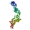
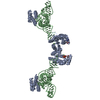
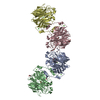
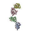
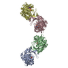
| Title | Crystal Structure of Myosin-1c tail in complex with Calmodulin | ||||||
 Components Components |
| ||||||
 Keywords Keywords | PROTEIN BINDING/CALCIUM-BINDING PROTEIN /  EF hand / PH domain / IQ motif / Myosin / Ca2+ signaling / Force sensing / Calcium binding / lipid binding / plasma membrane / cytoskeleton / PROTEIN BINDING-CALCIUM-BINDING PROTEIN complex EF hand / PH domain / IQ motif / Myosin / Ca2+ signaling / Force sensing / Calcium binding / lipid binding / plasma membrane / cytoskeleton / PROTEIN BINDING-CALCIUM-BINDING PROTEIN complex | ||||||
| Function / homology |  Function and homology information Function and homology informationstereocilium membrane / small GTPase binding => GO:0031267 / positive regulation of cellular response to insulin stimulus / B-WICH complex positively regulates rRNA expression / microfilament motor activity => GO:0000146 / : / positive regulation of vascular endothelial growth factor signaling pathway / stereocilium bundle / positive regulation of cell migration by vascular endothelial growth factor signaling pathway / Regulation of actin dynamics for phagocytic cup formation ...stereocilium membrane / small GTPase binding => GO:0031267 / positive regulation of cellular response to insulin stimulus / B-WICH complex positively regulates rRNA expression / microfilament motor activity => GO:0000146 / : / positive regulation of vascular endothelial growth factor signaling pathway / stereocilium bundle / positive regulation of cell migration by vascular endothelial growth factor signaling pathway / Regulation of actin dynamics for phagocytic cup formation / stereocilium / vesicle transport along actin filament / protein targeting to membrane / regulation of bicellular tight junction assembly / myosin complex / microfilament motor activity / positive regulation of actin filament polymerization / filamentous actin / microvillus / brush border / positive regulation of protein targeting to membrane / protein targeting / mRNA transport / lateral plasma membrane / phagocytic vesicle / nuclear pore / basal plasma membrane / actin filament organization / phospholipid binding / cytoplasmic vesicle membrane / ruffle membrane / cellular response to type II interferon / actin filament binding / actin cytoskeleton / actin binding / cytoplasmic vesicle / vesicle / nuclear body / calmodulin binding / positive regulation of cell migration / membrane raft / signaling receptor binding / calcium ion binding / nucleolus / ATP binding / membrane / nucleus / plasma membrane / cytosol / cytoplasmSimilarity search - Function | ||||||
| Biological species |  Mus musculus (house mouse)Xenopus laevis (African clawed frog) Mus musculus (house mouse)Xenopus laevis (African clawed frog) | ||||||
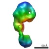
| Method | X-RAY DIFFRACTION / SYNCHROTRON / SAD / Resolution: 3.503 Å | ||||||
 Authors Authors | Lu, Q. / Li, J. / Ye, F. / Zhang, M. | ||||||
 Citation Citation | Journal: Nat.Struct.Mol.Biol. / Year: 2015 Title: Structure of myosin-1c tail bound to calmodulin provides insights into calcium-mediated conformational coupling. Authors: Lu, Q. / Li, J. / Ye, F. / Zhang, M. | ||||||
| History |
|
- Structure visualization
| Structure viewer | Molecule: MolmilJmol/JSmol |
|---|
- Downloads & links
-Download
| PDBx/mmCIF format | 4r8g.cif.gz | 153.9 KB | Display | PDBx/mmCIF format |
|---|---|---|---|---|
| PDB format | pdb4r8g.ent.gz | 119.3 KB | Display | PDB format |
| PDBx/mmJSON format | 4r8g.json.gz | Tree view | PDBx/mmJSON format | |
| Others |  Other downloads Other downloads |
-Validation report
| Arichive directory | https://data.pdbj.org/pub/pdb/validation_reports/r8/4r8gftp://data.pdbj.org/pub/pdb/validation_reports/r8/4r8g | HTTPS FTP |
|---|
-Related structure data
| Similar structure data |
|---|


-Links
 PDBj
PDBj


- Assembly
| Deposited unit | 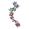
| ||||||||
|---|---|---|---|---|---|---|---|---|---|
| 1 |
| ||||||||
| Unit cell |
|
-Components
| #1: Protein | Mass: 38111.070 Da / Num. of mol.: 1 / Fragment: UNP residues 733-1063 Source method: isolated from a genetically manipulated source Source: (gene. exp.) Mus musculus (house mouse) / Gene: Myo1c / Production host:  Escherichia coli (E. coli) / References: UniProt: Q9WTI7 Escherichia coli (E. coli) / References: UniProt: Q9WTI7 | ||
|---|---|---|---|
| #2: Protein | / CaM Mass: 16721.350 Da / Num. of mol.: 3 Source method: isolated from a genetically manipulated source Source: (gene. exp.) Xenopus laevis (African clawed frog) / Gene: calm1, calm2 / Production host: Escherichia coli (E. coli) / References: UniProt: P62155, UniProt: P0DP33*PLUS#3: Chemical | Sulfate  Mass: 96.063 Da / Num. of mol.: 3 / Source method: obtained synthetically / Formula: SO4 Mass: 96.063 Da / Num. of mol.: 3 / Source method: obtained synthetically / Formula: SO4 |
-Experimental details
-Experiment
| Experiment | Method: X-RAY DIFFRACTION / Number of used crystals: 1 |
|---|
- Sample preparation
| Crystal | Density Matthews: 4.54 Å3/Da / Density % sol: 72.88 % |
|---|---|
| Crystal grow | Temperature: 289.3 K / Method: vapor diffusion, hanging drop / pH: 6.5 Details: 2.0M ammonium sulfate, 5% 1,4-dioxane, MES pH 6.5, VAPOR DIFFUSION, HANGING DROP, temperature 289.3K |
-Data collection
| Diffraction | Mean temperature: 100 K |
|---|---|
| Diffraction source | Source: SYNCHROTRON / Site: SSRF  / Beamline: BL17U / Wavelength: 0.9793 Å / Beamline: BL17U / Wavelength: 0.9793 Å |
| Detector | Type: ADSC QUANTUM 315r / Detector: CCD / Date: Apr 30, 2012 |
| Radiation | Monochromator: double crystal / Protocol: SINGLE WAVELENGTH / Monochromatic (M) / Laue (L): M / Scattering type: x-ray |
| Radiation wavelength | Wavelength: 0.9793 Å / Relative weight: 1 |
| Reflection | Resolution: 3.5→50 Å / Num. obs: 21359 / Biso Wilson estimate: 107.11 Å2 |
- Processing
| Software |
| |||||||||||||||||||||||||||||||||||||||||||||
|---|---|---|---|---|---|---|---|---|---|---|---|---|---|---|---|---|---|---|---|---|---|---|---|---|---|---|---|---|---|---|---|---|---|---|---|---|---|---|---|---|---|---|---|---|---|---|
| Refinement | Method to determine structure: SAD / Resolution: 3.503→45.59 Å / SU ML: 0.47 / σ(F): 1.34 / Phase error: 31.73 / Stereochemistry target values: ML
| |||||||||||||||||||||||||||||||||||||||||||||
| Solvent computation | Shrinkage radii: 0.9 Å / VDW probe radii: 1.11 Å / Solvent model: FLAT BULK SOLVENT MODEL | |||||||||||||||||||||||||||||||||||||||||||||
| Refinement step | Cycle: LAST / Resolution: 3.503→45.59 Å
| |||||||||||||||||||||||||||||||||||||||||||||
| Refine LS restraints |
| |||||||||||||||||||||||||||||||||||||||||||||
| LS refinement shell | Refine-ID: X-RAY DIFFRACTION / Total num. of bins used: 8 / % reflection obs: 100 %
|
