 Movie
Movie Controller

[English] 日本語
 Yorodumi
- PDB-4qx1: Cry3A Toxin structure obtained by Serial Femtosecond Crystallogra... -
+ Open data

- Basic information
| Entry | Database: PDB / ID: 4qx1 | ||||||
|---|---|---|---|---|---|---|---|
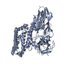
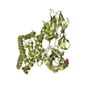
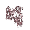
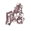
| Title | Cry3A Toxin structure obtained by Serial Femtosecond Crystallography from in vivo grown crystals isolated from Bacillus thuringiensis and data processed with the CrystFEL software suite | ||||||
 Components Components | Pesticidal crystal protein cry3Aa | ||||||
 Keywords Keywords |  TOXIN / in vivo crystals / microcrystals / serial femtosecond crystallography / SFX / LCLS / X-ray Free-electron laser / insecticidal toxin TOXIN / in vivo crystals / microcrystals / serial femtosecond crystallography / SFX / LCLS / X-ray Free-electron laser / insecticidal toxin | ||||||
| Function / homology |  Function and homology information Function and homology informationsymbiont-mediated killing of host cell / sporulation resulting in formation of a cellular spore / toxin activity / signaling receptor bindingSimilarity search - Function | ||||||
| Biological species |  Bacillus thuringiensis serovar tenebrionis (bacteria) Bacillus thuringiensis serovar tenebrionis (bacteria) | ||||||
| Method | X-RAY DIFFRACTION / FREE ELECTRON LASER / FOURIER SYNTHESIS / Resolution: 2.8 Å | ||||||
 Authors Authors | Sawaya, M.R. / Cascio, D. / Gingery, M. / Rodriguez, J. / Goldschmidt, L. / Colletier, J.-P. / Messerschmidt, M. / Boutet, S. / Koglin, J.E. / Williams, G.J. ...Sawaya, M.R. / Cascio, D. / Gingery, M. / Rodriguez, J. / Goldschmidt, L. / Colletier, J.-P. / Messerschmidt, M. / Boutet, S. / Koglin, J.E. / Williams, G.J. / Brewster, A.S. / Nass, K. / Hattne, J. / Botha, S. / Doak, R.B. / Shoeman, R.L. / DePonte, D.P. / Park, H.-W. / Federici, B.A. / Sauter, N.K. / Schlichting, I. / Eisenberg, D. | ||||||
 Citation Citation | Journal: Proc.Natl.Acad.Sci.USA / Year: 2014 Title: Protein crystal structure obtained at 2.9 angstrom resolution from injecting bacterial cells into an X-ray free-electron laser beam. Authors: Sawaya, M.R. / Cascio, D. / Gingery, M. / Rodriguez, J. / Goldschmidt, L. / Colletier, J.P. / Messerschmidt, M.M. / Boutet, S. / Koglin, J.E. / Williams, G.J. / Brewster, A.S. / Nass, K. / ...Authors: Sawaya, M.R. / Cascio, D. / Gingery, M. / Rodriguez, J. / Goldschmidt, L. / Colletier, J.P. / Messerschmidt, M.M. / Boutet, S. / Koglin, J.E. / Williams, G.J. / Brewster, A.S. / Nass, K. / Hattne, J. / Botha, S. / Doak, R.B. / Shoeman, R.L. / DePonte, D.P. / Park, H.W. / Federici, B.A. / Sauter, N.K. / Schlichting, I. / Eisenberg, D.S. | ||||||
| History |
|
- Structure visualization
| Structure viewer | Molecule: MolmilJmol/JSmol |
|---|
- Downloads & links
-Download
| PDBx/mmCIF format | 4qx1.cif.gz | 243.8 KB | Display | PDBx/mmCIF format |
|---|---|---|---|---|
| PDB format | pdb4qx1.ent.gz | 198 KB | Display | PDB format |
| PDBx/mmJSON format | 4qx1.json.gz | Tree view | PDBx/mmJSON format | |
| Others |  Other downloads Other downloads |
-Validation report
| Arichive directory | https://data.pdbj.org/pub/pdb/validation_reports/qx/4qx1ftp://data.pdbj.org/pub/pdb/validation_reports/qx/4qx1 | HTTPS FTP |
|---|
-Related structure data
| Related structure data | 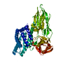 4qx0C  4qx2C  4qx3C 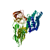 1dlcS C: citing same article ( S: Starting model for refinement |
|---|---|
| Similar structure data |


-Links
 PDBj
PDBj- Assembly
| Deposited unit | 
| ||||||||
|---|---|---|---|---|---|---|---|---|---|
| 1 |
| ||||||||
| Unit cell |
|
-Components
| #1: Protein | Mass: 66602.039 Da / Num. of mol.: 1 / Fragment: UNP residues 58-644 Source method: isolated from a genetically manipulated source Source: (gene. exp.) Bacillus thuringiensis serovar tenebrionis (bacteria)Strain: tenebrionis / Gene: bt13, cry3A, cry3Aa, cryC, cryIIIa, cryIIIA(a) / Plasmid: pPFT3As Production host: Bacillus thuringiensis subsp. israelensis (bacteria)Strain (production host): 4Q7 / References: UniProt: P0A379 |
|---|
-Experimental details
-Experiment
| Experiment | Method: X-RAY DIFFRACTION / Number of used crystals: 1 |
|---|
- Sample preparation
| Crystal | Density Matthews: 3.14 Å3/Da / Density % sol: 60.88 % |
|---|---|
| Crystal grow | Temperature: 303 K / Method: in vivo crystallization Details: Cry3A crystallized within the bacterial cell, IN VIVO CRYSTALLIZATION, temperature 303K |
-Data collection
| Diffraction | Mean temperature: 298 K |
|---|---|
| Diffraction source | Source: FREE ELECTRON LASER / Site: SLAC LCLS  / Beamline: CXI / Wavelength: 1.456 Å / Beamline: CXI / Wavelength: 1.456 Å |
| Detector | Type: Cornell-SLAC Pixel Array Detector (CSPAD) / Detector: PIXEL / Date: Mar 11, 2013 |
| Radiation | Monochromator: no monochromator, FEL beam with 20-30 eV bandwidth Protocol: SINGLE WAVELENGTH / Monochromatic (M) / Laue (L): M / Scattering type: x-ray |
| Radiation wavelength | Wavelength: 1.456 Å / Relative weight: 1 |
| Reflection | Resolution: 2.8→88.6 Å / Num. all: 21038 / Num. obs: 21038 / % possible obs: 100 % / Redundancy: 1128 % / Biso Wilson estimate: 80.64 Å2 / Rmerge(I) obs: 0.159 / Net I/σ(I): 9.5 |
| Reflection shell | Resolution: 2.8→2.9 Å / Redundancy: 691.5 % / Rmerge(I) obs: 0.412 / Mean I/σ(I) obs: 1.6 / % possible all: 100 |
- Processing
| Software |
| ||||||||||||||||||||||||||||||||||||||||||||||||||||||||||||||||||||||||||||||||||||||||||||||||||||||||||||||||||
|---|---|---|---|---|---|---|---|---|---|---|---|---|---|---|---|---|---|---|---|---|---|---|---|---|---|---|---|---|---|---|---|---|---|---|---|---|---|---|---|---|---|---|---|---|---|---|---|---|---|---|---|---|---|---|---|---|---|---|---|---|---|---|---|---|---|---|---|---|---|---|---|---|---|---|---|---|---|---|---|---|---|---|---|---|---|---|---|---|---|---|---|---|---|---|---|---|---|---|---|---|---|---|---|---|---|---|---|---|---|---|---|---|---|---|---|
| Refinement | Method to determine structure: FOURIER SYNTHESIS Starting model: PDB ENTRY 1DLC Resolution: 2.8→44.29 Å / Cor.coef. Fo:Fc: 0.9466 / Cor.coef. Fo:Fc free: 0.9377 / SU R Cruickshank DPI: 0.695 / Cross valid method: THROUGHOUT / σ(F): 0 / SU R Blow DPI: 0.624 / SU Rfree Blow DPI: 0.252 / SU Rfree Cruickshank DPI: 0.26 / Stereochemistry target values: Engh & Huber
| ||||||||||||||||||||||||||||||||||||||||||||||||||||||||||||||||||||||||||||||||||||||||||||||||||||||||||||||||||
| Displacement parameters | Biso mean: 75.68 Å2
| ||||||||||||||||||||||||||||||||||||||||||||||||||||||||||||||||||||||||||||||||||||||||||||||||||||||||||||||||||
| Refine analyze | Luzzati coordinate error obs: 0.41 Å | ||||||||||||||||||||||||||||||||||||||||||||||||||||||||||||||||||||||||||||||||||||||||||||||||||||||||||||||||||
| Refinement step | Cycle: LAST / Resolution: 2.8→44.29 Å
| ||||||||||||||||||||||||||||||||||||||||||||||||||||||||||||||||||||||||||||||||||||||||||||||||||||||||||||||||||
| Refine LS restraints |
| ||||||||||||||||||||||||||||||||||||||||||||||||||||||||||||||||||||||||||||||||||||||||||||||||||||||||||||||||||
| LS refinement shell | Resolution: 2.8→2.94 Å / Total num. of bins used: 11
| ||||||||||||||||||||||||||||||||||||||||||||||||||||||||||||||||||||||||||||||||||||||||||||||||||||||||||||||||||
| Refinement TLS params. | Method: refined / Origin x: 36.3377 Å / Origin y: 42.6468 Å / Origin z: 46.8855 Å
| ||||||||||||||||||||||||||||||||||||||||||||||||||||||||||||||||||||||||||||||||||||||||||||||||||||||||||||||||||
| Refinement TLS group | Selection details: { A|* } |
