 Movie
Movie Controller

[English] 日本語
 Yorodumi
- PDB-4qt8: Crystal Structure of RON Sema-PSI-IPT1 extracellular domains in c... -
+ Open data

- Basic information
| Entry | Database: PDB / ID: 4qt8 | |||||||||
|---|---|---|---|---|---|---|---|---|---|---|
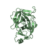
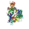
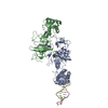
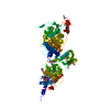
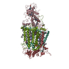
| Title | Crystal Structure of RON Sema-PSI-IPT1 extracellular domains in complex with MSP beta-chain | |||||||||
 Components Components |
| |||||||||
 Keywords Keywords | HYDROLASE/SIGNALING PROTEIN / Growth Factor receptor/Growth factor / Receptor-ligand complex / RON receptor tyrosine kinase /  Macrophage Stimulating Protein / HYDROLASE-SIGNALING PROTEIN complex Macrophage Stimulating Protein / HYDROLASE-SIGNALING PROTEIN complex | |||||||||
| Function / homology |  Function and homology information Function and homology informationSignaling by MST1 / macrophage colony-stimulating factor receptor activity / regulation of cAMP-dependent protein kinase activity / vacuole / regulation of receptor signaling pathway via JAK-STAT / single fertilization / negative regulation of gluconeogenesis / phagocytosis / stress fiber / transmembrane receptor protein tyrosine kinase activity ...Signaling by MST1 / macrophage colony-stimulating factor receptor activity / regulation of cAMP-dependent protein kinase activity / vacuole / regulation of receptor signaling pathway via JAK-STAT / single fertilization / negative regulation of gluconeogenesis / phagocytosis / stress fiber / transmembrane receptor protein tyrosine kinase activity / response to virus / positive regulation of MAP kinase activity / defense response / receptor tyrosine kinase binding / receptor protein-tyrosine kinase / cell surface receptor protein tyrosine kinase signaling pathway / cell migration / nervous system development / collagen-containing extracellular matrix / positive regulation of phosphatidylinositol 3-kinase/protein kinase B signal transduction / receptor complex / phosphorylation / innate immune response / positive regulation of cell population proliferation / enzyme binding / cell surface / signal transduction / proteolysis / extracellular space / extracellular region / ATP binding / plasma membraneSimilarity search - Function | |||||||||
| Biological species |  Homo sapiens (human) Homo sapiens (human) | |||||||||
| Method | X-RAY DIFFRACTION / SYNCHROTRON / MOLECULAR REPLACEMENT / Resolution: 3 Å | |||||||||
 Authors Authors | Herzberg, O. / Chao, K.L. | |||||||||
 Citation Citation | Journal: J.Biol.Chem. / Year: 2014 Title: Structural basis for the binding specificity of human Recepteur d'Origine Nantais (RON) receptor tyrosine kinase to macrophage-stimulating protein. Authors: Chao, K.L. / Gorlatova, N.V. / Eisenstein, E. / Herzberg, O. | |||||||||
| History |
|
- Structure visualization
| Structure viewer | Molecule: MolmilJmol/JSmol |
|---|
- Downloads & links
-Download
| PDBx/mmCIF format | 4qt8.cif.gz | 332.8 KB | Display | PDBx/mmCIF format |
|---|---|---|---|---|
| PDB format | pdb4qt8.ent.gz | 262.4 KB | Display | PDB format |
| PDBx/mmJSON format | 4qt8.json.gz | Tree view | PDBx/mmJSON format | |
| Others |  Other downloads Other downloads |
-Validation report
| Arichive directory | https://data.pdbj.org/pub/pdb/validation_reports/qt/4qt8ftp://data.pdbj.org/pub/pdb/validation_reports/qt/4qt8 | HTTPS FTP |
|---|
-Related structure data


-Links
 PDBj
PDBj


- Assembly
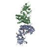
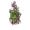
| Deposited unit | 
| ||||||||||||||||||||||||||||||||||||||||||||||||||||||||||||||||||||
|---|---|---|---|---|---|---|---|---|---|---|---|---|---|---|---|---|---|---|---|---|---|---|---|---|---|---|---|---|---|---|---|---|---|---|---|---|---|---|---|---|---|---|---|---|---|---|---|---|---|---|---|---|---|---|---|---|---|---|---|---|---|---|---|---|---|---|---|---|---|
| 1 | 
| ||||||||||||||||||||||||||||||||||||||||||||||||||||||||||||||||||||
| 2 | 
| ||||||||||||||||||||||||||||||||||||||||||||||||||||||||||||||||||||
| Unit cell |
| ||||||||||||||||||||||||||||||||||||||||||||||||||||||||||||||||||||
| Noncrystallographic symmetry (NCS) | NCS domain:
NCS domain segments: Component-ID: 0 / Refine code: 0
NCS ensembles :
|
-Components
| #1: Protein | Mass: 71987.438 Da / Num. of mol.: 2 Fragment: extracellular Sema-PSI-IPT1 domains (UNP residues 25-683) Mutation: R306L, R307V, R308P, A311S, R322Q Source method: isolated from a genetically manipulated source Source: (gene. exp.) Homo sapiens (human) / Gene: MST1R, PTK8, RON / Plasmid: pMT/BiP/V5-His / Production host:  Drosophila melanogaster (fruit fly) / Strain (production host): S2 Drosophila melanogaster (fruit fly) / Strain (production host): S2References: UniProt: Q04912, receptor protein-tyrosine kinase#2: Protein | Mass: 28336.740 Da / Num. of mol.: 2 / Fragment: beta chain (UNP residues 465-711) / Mutation: C672S Source method: isolated from a genetically manipulated source Source: (gene. exp.) Homo sapiens (human) / Gene: D3F15S2, DNF15S2, HGFL, MSI1, MST1 / Plasmid: pMT/BiP/V5-His / Production host: Drosophila melanogaster (fruit fly) / Strain (production host): S2 / References: UniProt: P26927#3: Polysaccharide | beta-D-mannopyranose-(1-4)-2-acetamido-2-deoxy-beta-D-glucopyranose-(1-4)-2-acetamido-2-deoxy-beta- ...beta-D-mannopyranose-(1-4)-2-acetamido-2-deoxy-beta-D-glucopyranose-(1-4)-2-acetamido-2-deoxy-beta-D-glucopyranose | / Mass: 586.542 Da / Num. of mol.: 1Source method: isolated from a genetically manipulated source #4: Polysaccharide | 2-acetamido-2-deoxy-beta-D-glucopyranose-(1-4)-2-acetamido-2-deoxy-beta-D-glucopyranose | / Mass: 424.401 Da / Num. of mol.: 1Source method: isolated from a genetically manipulated source |
|---|
-Experimental details
-Experiment
| Experiment | Method: X-RAY DIFFRACTION / Number of used crystals: 1 |
|---|
- Sample preparation
| Crystal | Density Matthews: 2.49 Å3/Da / Density % sol: 50.54 % |
|---|---|
| Crystal grow | Temperature: 298 K / Method: vapor diffusion, sitting drop / pH: 8.5 Details: 0.1 M Tris-HCl, pH 8.5, 20% w/v PEG4000, 8% v/v isopropanol, 4% v/v polypropylene glycol 400, VAPOR DIFFUSION, SITTING DROP, temperature 298K |
-Data collection
| Diffraction | Mean temperature: 100 K | |||||||||||||||
|---|---|---|---|---|---|---|---|---|---|---|---|---|---|---|---|---|
| Diffraction source | Source: SYNCHROTRON / Site: APS  / Beamline: 23-ID-B / Wavelength: 1.0332 Å / Beamline: 23-ID-B / Wavelength: 1.0332 Å | |||||||||||||||
| Detector | Type: MARMOSAIC 300 mm CCD / Detector: CCD / Date: Nov 8, 2010 / Details: mirrors | |||||||||||||||
| Radiation | Monochromator: Double crystal cryo-cooled Si(111) / Protocol: SINGLE WAVELENGTH / Monochromatic (M) / Laue (L): M / Scattering type: x-ray | |||||||||||||||
| Radiation wavelength | Wavelength: 1.0332 Å / Relative weight: 1 | |||||||||||||||
| Reflection twin |
| |||||||||||||||
| Reflection | Resolution: 2.996→146.96 Å / Num. all: 39670 / Num. obs: 38960 / % possible obs: 91.8 % / Observed criterion σ(F): 0 / Observed criterion σ(I): -3 / Redundancy: 2.5 % / Rmerge(I) obs: 0.202 / Net I/σ(I): 4.4 | |||||||||||||||
| Reflection shell | Highest resolution: 2.996 Å |
- Processing
| Software |
| ||||||||||||||||||||||||||||||||||||||||||||||||||||||||||||||||||||||||||||||||||||||||||||||||||||||||||||||||||||||||||||||||||||||||||||||||||||||||||||||||||||||||||||||||||||||
|---|---|---|---|---|---|---|---|---|---|---|---|---|---|---|---|---|---|---|---|---|---|---|---|---|---|---|---|---|---|---|---|---|---|---|---|---|---|---|---|---|---|---|---|---|---|---|---|---|---|---|---|---|---|---|---|---|---|---|---|---|---|---|---|---|---|---|---|---|---|---|---|---|---|---|---|---|---|---|---|---|---|---|---|---|---|---|---|---|---|---|---|---|---|---|---|---|---|---|---|---|---|---|---|---|---|---|---|---|---|---|---|---|---|---|---|---|---|---|---|---|---|---|---|---|---|---|---|---|---|---|---|---|---|---|---|---|---|---|---|---|---|---|---|---|---|---|---|---|---|---|---|---|---|---|---|---|---|---|---|---|---|---|---|---|---|---|---|---|---|---|---|---|---|---|---|---|---|---|---|---|---|---|---|
| Refinement | Method to determine structure: MOLECULAR REPLACEMENT Starting model: PDB ENTRIES 4FWW & 2ASU Resolution: 3→19.98 Å / Cor.coef. Fo:Fc: 0.876 / Cor.coef. Fo:Fc free: 0.818 / SU B: 13.688 / SU ML: 0.275 / Cross valid method: THROUGHOUT / ESU R Free: 0.116 / Stereochemistry target values: MAXIMUM LIKELIHOOD / Details: HYDROGENS HAVE BEEN ADDED IN THE RIDING POSITIONS
| ||||||||||||||||||||||||||||||||||||||||||||||||||||||||||||||||||||||||||||||||||||||||||||||||||||||||||||||||||||||||||||||||||||||||||||||||||||||||||||||||||||||||||||||||||||||
| Solvent computation | Ion probe radii: 0.8 Å / Shrinkage radii: 0.8 Å / VDW probe radii: 1.2 Å / Solvent model: MASK | ||||||||||||||||||||||||||||||||||||||||||||||||||||||||||||||||||||||||||||||||||||||||||||||||||||||||||||||||||||||||||||||||||||||||||||||||||||||||||||||||||||||||||||||||||||||
| Displacement parameters | Biso mean: 51.191 Å2
| ||||||||||||||||||||||||||||||||||||||||||||||||||||||||||||||||||||||||||||||||||||||||||||||||||||||||||||||||||||||||||||||||||||||||||||||||||||||||||||||||||||||||||||||||||||||
| Refinement step | Cycle: LAST / Resolution: 3→19.98 Å
| ||||||||||||||||||||||||||||||||||||||||||||||||||||||||||||||||||||||||||||||||||||||||||||||||||||||||||||||||||||||||||||||||||||||||||||||||||||||||||||||||||||||||||||||||||||||
| Refine LS restraints |
|