 Movie
Movie Controller

[English] 日本語
 Yorodumi
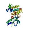
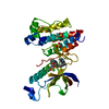
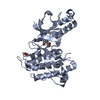
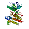
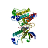
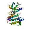
- PDB-4qqc: Crystal Structure of FGF Receptor (FGFR) 4 Kinase Domain in Compl... -
+ Open data

- Basic information
| Entry | Database: PDB / ID: 4qqc | ||||||
|---|---|---|---|---|---|---|---|
| Title | Crystal Structure of FGF Receptor (FGFR) 4 Kinase Domain in Complex with FIIN-2, an Irreversible Tyrosine Kinase Inhibitor Capable of Overcoming FGFR Kinase Gate-Keeper Mutations | ||||||
 Components Components | Fibroblast growth factor receptor 4 | ||||||
 Keywords Keywords | Transferase/transferase inhibitor / Kinase Domain Fold / Cell Signaling / Phosphotransferase / Plasmamembrane / Transferase-transferase inhibitor complex | ||||||
| Function / homology |  Function and homology information Function and homology informationFGFR4 mutant receptor activation / betaKlotho-mediated ligand binding / regulation of extracellular matrix disassembly / phosphate ion homeostasis / regulation of bile acid biosynthetic process / FGFR4 ligand binding and activation / fibroblast growth factor receptor activity / Phospholipase C-mediated cascade; FGFR4 / positive regulation of DNA biosynthetic process / positive regulation of catalytic activity ...FGFR4 mutant receptor activation / betaKlotho-mediated ligand binding / regulation of extracellular matrix disassembly / phosphate ion homeostasis / regulation of bile acid biosynthetic process / FGFR4 ligand binding and activation / fibroblast growth factor receptor activity / Phospholipase C-mediated cascade; FGFR4 / positive regulation of DNA biosynthetic process / positive regulation of catalytic activity / fibroblast growth factor binding / PI-3K cascade:FGFR4 / positive regulation of proteolysis / regulation of lipid metabolic process / PI3K Cascade / fibroblast growth factor receptor signaling pathway / SHC-mediated cascade:FGFR4 / Signaling by FGFR4 in disease / transport vesicle / FRS-mediated FGFR4 signaling / cholesterol homeostasis / Negative regulation of FGFR4 signaling / receptor protein-tyrosine kinase / peptidyl-tyrosine phosphorylation / cell surface receptor protein tyrosine kinase signaling pathway / Constitutive Signaling by Aberrant PI3K in Cancer / cell migration / glucose homeostasis / PIP3 activates AKT signaling / heparin binding / PI5P, PP2A and IER3 Regulate PI3K/AKT Signaling / RAF/MAP kinase cascade / protein autophosphorylation / positive regulation of ERK1 and ERK2 cascade / receptor complex / endosome / positive regulation of cell population proliferation / positive regulation of gene expression / Golgi apparatus / endoplasmic reticulum / extracellular region / ATP binding / plasma membraneSimilarity search - Function | ||||||
| Biological species |  Homo sapiens (human) Homo sapiens (human) | ||||||
| Method | X-RAY DIFFRACTION / SYNCHROTRON / MOLECULAR REPLACEMENT / Resolution: 2.4 Å | ||||||
 Authors Authors | Huang, Z. / Mohammadi, M. | ||||||
 Citation Citation | Journal: Proc.Natl.Acad.Sci.USA / Year: 2014 Title: Development of covalent inhibitors that can overcome resistance to first-generation FGFR kinase inhibitors. Authors: Tan, L. / Wang, J. / Tanizaki, J. / Huang, Z. / Aref, A.R. / Rusan, M. / Zhu, S.J. / Zhang, Y. / Ercan, D. / Liao, R.G. / Capelletti, M. / Zhou, W. / Hur, W. / Kim, N. / Sim, T. / Gaudet, S. ...Authors: Tan, L. / Wang, J. / Tanizaki, J. / Huang, Z. / Aref, A.R. / Rusan, M. / Zhu, S.J. / Zhang, Y. / Ercan, D. / Liao, R.G. / Capelletti, M. / Zhou, W. / Hur, W. / Kim, N. / Sim, T. / Gaudet, S. / Barbie, D.A. / Yeh, J.R. / Yun, C.H. / Hammerman, P.S. / Mohammadi, M. / Janne, P.A. / Gray, N.S. | ||||||
| History |
|
- Structure visualization
| Structure viewer | Molecule: MolmilJmol/JSmol |
|---|
- Downloads & links
-Download
| PDBx/mmCIF format | 4qqc.cif.gz | 72.4 KB | Display | PDBx/mmCIF format |
|---|---|---|---|---|
| PDB format | pdb4qqc.ent.gz | 52 KB | Display | PDB format |
| PDBx/mmJSON format | 4qqc.json.gz | Tree view | PDBx/mmJSON format | |
| Others |  Other downloads Other downloads |
-Validation report
| Arichive directory | https://data.pdbj.org/pub/pdb/validation_reports/qq/4qqcftp://data.pdbj.org/pub/pdb/validation_reports/qq/4qqc | HTTPS FTP |
|---|
-Related structure data
| Related structure data | 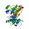 4r5sC  4r6vC  2psqS C: citing same article ( S: Starting model for refinement |
|---|---|
| Similar structure data |


-Links
 PDBj
PDBj


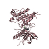
- Assembly
| Deposited unit | 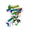
| ||||||||
|---|---|---|---|---|---|---|---|---|---|
| 1 |
| ||||||||
| Unit cell |
|
-Components
| #1: Protein | / FGFR-4 Mass: 36159.574 Da / Num. of mol.: 1 / Fragment: Kinase domain Of FGF Receptor 4 / Mutation: R669E Source method: isolated from a genetically manipulated source Source: (gene. exp.) Homo sapiens (human) / Gene: FGFR4, JTK2, TKF / Production host:  Escherichia coli (E. coli) Escherichia coli (E. coli)References: UniProt: P22455, receptor protein-tyrosine kinase |
|---|---|
| #2: Chemical | ChemComp-37O / 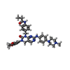  Mass: 636.743 Da / Num. of mol.: 1 / Source method: obtained synthetically / Formula: C35H40N8O4 Mass: 636.743 Da / Num. of mol.: 1 / Source method: obtained synthetically / Formula: C35H40N8O4 |
| #3: Chemical | ChemComp-SO4 / Sulfate  Mass: 96.063 Da / Num. of mol.: 1 / Source method: obtained synthetically / Formula: SO4 Mass: 96.063 Da / Num. of mol.: 1 / Source method: obtained synthetically / Formula: SO4 |
| #4: Water | ChemComp-HOH / Water Mass: 18.015 Da / Num. of mol.: 41 / Source method: isolated from a natural source / Formula: H2O Mass: 18.015 Da / Num. of mol.: 41 / Source method: isolated from a natural source / Formula: H2O |
-Experimental details
-Experiment
| Experiment | Method: X-RAY DIFFRACTION / Number of used crystals: 1 |
|---|
- Sample preparation
| Crystal | Density Matthews: 2.57 Å3/Da / Density % sol: 52.23 % |
|---|---|
| Crystal grow | Temperature: 291 K / Method: vapor diffusion, hanging drop / pH: 7.5 Details: 0.1 M HEPES, 1.0 1.2 M (NH4)2SO4, 10 mM Yttrium (III) chloride hexahydrate, pH 7.5, VAPOR DIFFUSION, HANGING DROP, temperature 291K |
-Data collection
| Diffraction | Mean temperature: 100 K |
|---|---|
| Diffraction source | Source: SYNCHROTRON / Site: NSLS  / Beamline: X4C / Wavelength: 0.979 Å / Beamline: X4C / Wavelength: 0.979 Å |
| Detector | Type: MAR CCD 165 mm / Detector: CCD / Date: Mar 14, 2014 |
| Radiation | Monochromator: Si 111 Channel / Protocol: SINGLE WAVELENGTH / Monochromatic (M) / Laue (L): M / Scattering type: x-ray |
| Radiation wavelength | Wavelength: 0.979 Å / Relative weight: 1 |
| Reflection | Resolution: 2.35→50 Å / Num. all: 15298 / Num. obs: 15298 / % possible obs: 100 % / Observed criterion σ(F): 0 / Observed criterion σ(I): 0 / Redundancy: 11.3 % / Rmerge(I) obs: 0.106 / Rsym value: 0.106 / Net I/σ(I): 29.7 |
| Reflection shell | Resolution: 2.35→2.39 Å / % possible all: 99.6 |
- Processing
| Software |
| |||||||||||||||||||||||||||||||||||||||||||||||||||||||||||||||||||||||||||||
|---|---|---|---|---|---|---|---|---|---|---|---|---|---|---|---|---|---|---|---|---|---|---|---|---|---|---|---|---|---|---|---|---|---|---|---|---|---|---|---|---|---|---|---|---|---|---|---|---|---|---|---|---|---|---|---|---|---|---|---|---|---|---|---|---|---|---|---|---|---|---|---|---|---|---|---|---|---|---|
| Refinement | Method to determine structure: MOLECULAR REPLACEMENT Starting model: pdb entry 2PSQ Resolution: 2.4→27.79 Å / SU ML: 0.25 / σ(F): 2.04 / Phase error: 20.52 / Stereochemistry target values: ML
| |||||||||||||||||||||||||||||||||||||||||||||||||||||||||||||||||||||||||||||
| Solvent computation | Shrinkage radii: 0.9 Å / VDW probe radii: 1.11 Å / Solvent model: FLAT BULK SOLVENT MODEL | |||||||||||||||||||||||||||||||||||||||||||||||||||||||||||||||||||||||||||||
| Refinement step | Cycle: LAST / Resolution: 2.4→27.79 Å
| |||||||||||||||||||||||||||||||||||||||||||||||||||||||||||||||||||||||||||||
| Refine LS restraints |
| |||||||||||||||||||||||||||||||||||||||||||||||||||||||||||||||||||||||||||||
| LS refinement shell |
| |||||||||||||||||||||||||||||||||||||||||||||||||||||||||||||||||||||||||||||
| Refinement TLS params. | Method: refined / Origin x: -22.674 Å / Origin y: 3.6359 Å / Origin z: -9.834 Å
| |||||||||||||||||||||||||||||||||||||||||||||||||||||||||||||||||||||||||||||
| Refinement TLS group | Selection details: all |
