 Movie
Movie Controller


+ Open data

- Basic information
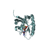
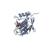
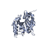
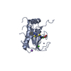
| Entry | Database: PDB / ID: 4qpn | ||||||
|---|---|---|---|---|---|---|---|
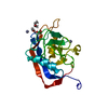
| Title | Crystal Structure of Human Methyltransferase-Like Protein 21B | ||||||
 Components Components | Protein-lysine methyltransferase METTL21B | ||||||
 Keywords Keywords |  TRANSFERASE / structural genomics / Structural Genomics Consortium / SGC TRANSFERASE / structural genomics / Structural Genomics Consortium / SGC | ||||||
| Function / homology |  Function and homology information Function and homology informationpeptidyl-lysine methylation / intracellular anatomical structure / protein-lysine N-methyltransferase activity / heat shock protein binding / Transferases; Transferring one-carbon groups; Methyltransferases / methyltransferase activity / centrosome / protein-containing complex / nucleoplasm / cytosol / cytoplasmSimilarity search - Function | ||||||
| Biological species |  Homo sapiens (human) Homo sapiens (human) | ||||||
| Method | X-RAY DIFFRACTION / SYNCHROTRON / MOLECULAR REPLACEMENT / molecular replacement / Resolution: 1.25 Å | ||||||
 Authors Authors | Tempel, W. / Hong, B.S. / Seitova, A. / He, H. / Li, Y. / Graslund, S. / Arrowsmith, C.H. / Edwards, A.M. / Brown, P.J. / Structural Genomics Consortium (SGC) | ||||||
 Citation Citation | Journal: TO BE PUBLISHED Title: Crystal Structure of Human Methyltransferase-Like Protein 21B Authors: Tempel, W. / Hong, B.S. / Seitova, A. / He, H. / Li, Y. / Graslund, S. / Arrowsmith, C.H. / Edwards, A.M. / Brown, P.J. / Structural Genomics Consortium (SGC) | ||||||
| History |
|
- Structure visualization
| Structure viewer | Molecule: MolmilJmol/JSmol |
|---|
- Downloads & links
-Download
| PDBx/mmCIF format | 4qpn.cif.gz | 108.8 KB | Display | PDBx/mmCIF format |
|---|---|---|---|---|
| PDB format | pdb4qpn.ent.gz | 81.8 KB | Display | PDB format |
| PDBx/mmJSON format | 4qpn.json.gz | Tree view | PDBx/mmJSON format | |
| Others |  Other downloads Other downloads |
-Validation report
| Arichive directory | https://data.pdbj.org/pub/pdb/validation_reports/qp/4qpnftp://data.pdbj.org/pub/pdb/validation_reports/qp/4qpn | HTTPS FTP |
|---|
-Related structure data
| Related structure data |  4lecS S: Starting model for refinement |
|---|---|
| Similar structure data |


-Links
 PDBj
PDBj- Assembly
| Deposited unit | 
| ||||||||
|---|---|---|---|---|---|---|---|---|---|
| 1 |
| ||||||||
| Unit cell |
|
-Components
| #1: Protein | Mass: 24994.111 Da / Num. of mol.: 1 Source method: isolated from a genetically manipulated source Source: (gene. exp.) Homo sapiens (human) / Gene: METTL21B, FAM119B, HCA557A / Plasmid: pFBOH-MHL / Production host:   Spodoptera frugiperda (fall armyworm) / Strain (production host): SF9 Spodoptera frugiperda (fall armyworm) / Strain (production host): SF9References: UniProt: Q96AZ1, Transferases; Transferring one-carbon groups; Methyltransferases | ||||
|---|---|---|---|---|---|
| #2: Chemical | ChemComp-SAH / S-Adenosyl-L-homocysteine  Type: L-peptide linking / Mass: 384.411 Da / Num. of mol.: 1 / Source method: obtained synthetically / Formula: C14H20N6O5S Type: L-peptide linking / Mass: 384.411 Da / Num. of mol.: 1 / Source method: obtained synthetically / Formula: C14H20N6O5S | ||||
| #3: Chemical | Sulfate  Mass: 96.063 Da / Num. of mol.: 2 / Source method: obtained synthetically / Formula: SO4 Mass: 96.063 Da / Num. of mol.: 2 / Source method: obtained synthetically / Formula: SO4#4: Chemical | ChemComp-UNX /   Num. of mol.: 14 / Source method: obtained synthetically Num. of mol.: 14 / Source method: obtained synthetically#5: Water | ChemComp-HOH / | Water Mass: 18.015 Da / Num. of mol.: 202 / Source method: isolated from a natural source / Formula: H2O Mass: 18.015 Da / Num. of mol.: 202 / Source method: isolated from a natural source / Formula: H2O |
-Experimental details
-Experiment
| Experiment | Method: X-RAY DIFFRACTION / Number of used crystals: 1 |
|---|
- Sample preparation
| Crystal | Density Matthews: 1.67 Å3/Da / Density % sol: 26.53 % |
|---|---|
| Crystal grow | Temperature: 293 K / Method: vapor diffusion Details: 20% PEG-3350, 0.2 M tri-lithium citrate. Protein sample was incubated with SAH overnight. Endopeptidase was added to the protein sample immediately prior to crystallization set up, vapor ...Details: 20% PEG-3350, 0.2 M tri-lithium citrate. Protein sample was incubated with SAH overnight. Endopeptidase was added to the protein sample immediately prior to crystallization set up, vapor diffusion, temperature 293K |
-Data collection
| Diffraction | Mean temperature: 100 K |
|---|---|
| Diffraction source | Source: SYNCHROTRON / Site: APS  / Beamline: 19-ID / Wavelength: 0.97915 Å / Beamline: 19-ID / Wavelength: 0.97915 Å |
| Detector | Type: adsc q315 / Detector: CCD / Date: Jun 12, 2014 |
| Radiation | Protocol: SINGLE WAVELENGTH / Monochromatic (M) / Laue (L): M / Scattering type: x-ray |
| Radiation wavelength | Wavelength: 0.97915 Å / Relative weight: 1 |
| Reflection | Resolution: 1.25→38.78 Å / Num. obs: 45170 / % possible obs: 100 % / Redundancy: 11.1 % / Rmerge(I) obs: 0.092 / Net I/σ(I): 19.6 |
| Reflection shell | Resolution: 1.25→1.27 Å / Redundancy: 10.8 % / Rmerge(I) obs: 0.956 / Mean I/σ(I) obs: 3 / Num. measured all: 23595 / Num. unique all: 2181 / % possible all: 100 |
-Phasing
| Phasing | Method: molecular replacement |
|---|
- Processing
| Software |
| ||||||||||||||||||||||||||||||||||||||||||||||||||||||||||||||||||||||||||||||||||||||||||
|---|---|---|---|---|---|---|---|---|---|---|---|---|---|---|---|---|---|---|---|---|---|---|---|---|---|---|---|---|---|---|---|---|---|---|---|---|---|---|---|---|---|---|---|---|---|---|---|---|---|---|---|---|---|---|---|---|---|---|---|---|---|---|---|---|---|---|---|---|---|---|---|---|---|---|---|---|---|---|---|---|---|---|---|---|---|---|---|---|---|---|---|
| Refinement | Method to determine structure: MOLECULAR REPLACEMENT Starting model: solved with data from isomorphous crystal. model based on pdb entry 4LEC. Resolution: 1.25→33.48 Å / Cor.coef. Fo:Fc: 0.98 / Cor.coef. Fo:Fc free: 0.97 / WRfactor Rfree: 0.1619 / WRfactor Rwork: 0.1185 / Occupancy max: 1 / Occupancy min: 0.3 / FOM work R set: 0.9047 / SU B: 1.482 / SU ML: 0.029 / SU R Cruickshank DPI: 0.0424 / SU Rfree: 0.0449 / Cross valid method: THROUGHOUT / σ(F): 0 / ESU R: 0.042 / ESU R Free: 0.045 / Stereochemistry target values: MAXIMUM LIKELIHOOD Details: Structure was solved and model was automatically built (ARP/WARP) using data from a nearly isomorphous crystal. COOT was used for interactive model building. Model geometry was evaluated with MOLPROBITY.
| ||||||||||||||||||||||||||||||||||||||||||||||||||||||||||||||||||||||||||||||||||||||||||
| Solvent computation | Ion probe radii: 0.8 Å / Shrinkage radii: 0.8 Å / VDW probe radii: 1.2 Å / Solvent model: MASK | ||||||||||||||||||||||||||||||||||||||||||||||||||||||||||||||||||||||||||||||||||||||||||
| Displacement parameters | Biso max: 65.27 Å2 / Biso mean: 12.1595 Å2 / Biso min: 4.05 Å2
| ||||||||||||||||||||||||||||||||||||||||||||||||||||||||||||||||||||||||||||||||||||||||||
| Refinement step | Cycle: LAST / Resolution: 1.25→33.48 Å
| ||||||||||||||||||||||||||||||||||||||||||||||||||||||||||||||||||||||||||||||||||||||||||
| Refine LS restraints |
| ||||||||||||||||||||||||||||||||||||||||||||||||||||||||||||||||||||||||||||||||||||||||||
| LS refinement shell | Resolution: 1.25→1.282 Å / Total num. of bins used: 20
|
