 Movie
Movie Controller


+ Open data

- Basic information
| Entry | Database: PDB / ID: 4qnc | ||||||
|---|---|---|---|---|---|---|---|
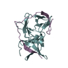
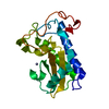
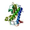
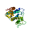
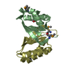
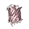
| Title | Crystal structure of a SemiSWEET in an occluded state | ||||||
 Components Components | chemical transport protein | ||||||
 Keywords Keywords |  MEMBRANE PROTEIN / transporter MEMBRANE PROTEIN / transporter | ||||||
| Function / homology |  Function and homology information Function and homology informationglucose transmembrane transporter activity / glucose transmembrane transport / protein homodimerization activity / membrane / plasma membraneSimilarity search - Function | ||||||
| Biological species |  Leptospira biflexa serovar Patoc (bacteria) Leptospira biflexa serovar Patoc (bacteria) | ||||||
| Method | X-RAY DIFFRACTION / SYNCHROTRON / MOLECULAR REPLACEMENT / Resolution: 2.388 Å | ||||||
 Authors Authors | Yan, X. / Yuyong, T. / Liang, F. / Perry, K. | ||||||
 Citation Citation | Journal: Nature / Year: 2014 Title: Structures of bacterial homologues of SWEET transporters in two distinct conformations. Authors: Xu, Y. / Tao, Y. / Cheung, L.S. / Fan, C. / Chen, L.Q. / Xu, S. / Perry, K. / Frommer, W.B. / Feng, L. | ||||||
| History |
|
- Structure visualization
| Structure viewer | Molecule: MolmilJmol/JSmol |
|---|
- Downloads & links
-Download
| PDBx/mmCIF format | 4qnc.cif.gz | 48.8 KB | Display | PDBx/mmCIF format |
|---|---|---|---|---|
| PDB format | pdb4qnc.ent.gz | 35.3 KB | Display | PDB format |
| PDBx/mmJSON format | 4qnc.json.gz | Tree view | PDBx/mmJSON format | |
| Others |  Other downloads Other downloads |
-Validation report
| Arichive directory | https://data.pdbj.org/pub/pdb/validation_reports/qn/4qncftp://data.pdbj.org/pub/pdb/validation_reports/qn/4qnc | HTTPS FTP |
|---|
-Related structure data


-Links
 PDBj
PDBj


- Assembly
| Deposited unit | 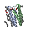
| ||||||||
|---|---|---|---|---|---|---|---|---|---|
| 1 |
| ||||||||
| Unit cell |
|
-Components
| #1: Protein | Mass: 10724.752 Da / Num. of mol.: 2 Source method: isolated from a genetically manipulated source Source: (gene. exp.) Leptospira biflexa serovar Patoc (bacteria)Strain: Patoc 1 / ATCC 23582 / Paris / Gene: LEPBI_I1613 / Production host: Escherichia coli (E. coli) / Strain (production host): BL21(DE3) / References: UniProt: B0SR19#2: Chemical | ChemComp-OLC / (   Mass: 356.540 Da / Num. of mol.: 4 / Source method: obtained synthetically / Formula: C21H40O4 Mass: 356.540 Da / Num. of mol.: 4 / Source method: obtained synthetically / Formula: C21H40O4#3: Chemical | ChemComp-MYS / Pentadecane  Mass: 212.415 Da / Num. of mol.: 4 / Source method: obtained synthetically / Formula: C15H32 Mass: 212.415 Da / Num. of mol.: 4 / Source method: obtained synthetically / Formula: C15H32#4: Water | ChemComp-HOH / | Water Mass: 18.015 Da / Num. of mol.: 14 / Source method: isolated from a natural source / Formula: H2O Mass: 18.015 Da / Num. of mol.: 14 / Source method: isolated from a natural source / Formula: H2O |
|---|
-Experimental details
-Experiment
| Experiment | Method: X-RAY DIFFRACTION / Number of used crystals: 11 |
|---|
- Sample preparation
| Crystal | Density Matthews: 2.09 Å3/Da / Density % sol: 41.05 % |
|---|---|
| Crystal grow | Temperature: 293 K / Method: lipidic cubic phase / pH: 7.5 Details: 100mM HEPES pH7.5, 150mM (NH4)2SO4, 10% PEG400, lipid cubic phase, temperature 293K |
-Data collection
| Diffraction |
| ||||||||||||||||||
|---|---|---|---|---|---|---|---|---|---|---|---|---|---|---|---|---|---|---|---|
| Diffraction source |
| ||||||||||||||||||
| Detector |
| ||||||||||||||||||
| Radiation |
| ||||||||||||||||||
| Radiation wavelength | Wavelength: 0.9794 Å / Relative weight: 1 | ||||||||||||||||||
| Reflection | Resolution: 2.388→50 Å / % possible obs: 95.5 % / Observed criterion σ(F): 0 / Observed criterion σ(I): -3 / Redundancy: 8 % / Rmerge(I) obs: 0.203 / Rsym value: 0.203 / Net I/σ(I): 11.1 | ||||||||||||||||||
| Reflection shell | Resolution: 2.4→2.44 Å / Redundancy: 2.3 % / Rmerge(I) obs: 0.556 / Mean I/σ(I) obs: 2 / Rsym value: 0.556 / % possible all: 79.4 |

- Processing
| Software |
| ||||||||||||||||||||||||
|---|---|---|---|---|---|---|---|---|---|---|---|---|---|---|---|---|---|---|---|---|---|---|---|---|---|
| Refinement | Method to determine structure: MOLECULAR REPLACEMENT / Resolution: 2.388→36.774 Å / SU ML: 0.36 / σ(F): 1.36 / Phase error: 32.42 / Stereochemistry target values: ML
| ||||||||||||||||||||||||
| Solvent computation | Shrinkage radii: 0.9 Å / VDW probe radii: 1.11 Å / Solvent model: FLAT BULK SOLVENT MODEL | ||||||||||||||||||||||||
| Refinement step | Cycle: LAST / Resolution: 2.388→36.774 Å
| ||||||||||||||||||||||||
| Refine LS restraints |
| ||||||||||||||||||||||||
| LS refinement shell |
|
