 Movie
Movie Controller


+ Open data

- Basic information
| Entry | Database: PDB / ID: 4qkv | ||||||
|---|---|---|---|---|---|---|---|
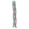
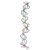
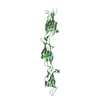
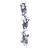
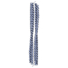
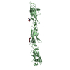
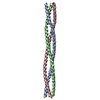
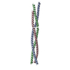
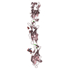
| Title | Crystal structure of the mouse cavin1 HR1 domain | ||||||
 Components Components | Polymerase I and transcript release factor | ||||||
 Keywords Keywords |  TRANSCRIPTION / coiled-coil / signalling / plasma membrane TRANSCRIPTION / coiled-coil / signalling / plasma membrane | ||||||
| Function / homology |  Function and homology information Function and homology informationRNA Polymerase I Transcription Termination / rRNA primary transcript binding / positive regulation of cell motility / termination of RNA polymerase I transcription / transcription initiation at RNA polymerase I promoter / rRNA transcription / protein secretion / caveola / membrane raft / intracellular membrane-bounded organelle ...RNA Polymerase I Transcription Termination / rRNA primary transcript binding / positive regulation of cell motility / termination of RNA polymerase I transcription / transcription initiation at RNA polymerase I promoter / rRNA transcription / protein secretion / caveola / membrane raft / intracellular membrane-bounded organelle / endoplasmic reticulum / protein-containing complex / mitochondrion / identical protein binding / nucleus / plasma membrane / cytosol / cytoplasmSimilarity search - Function | ||||||
| Biological species |  Mus musculus (house mouse) Mus musculus (house mouse) | ||||||
| Method | X-RAY DIFFRACTION / SYNCHROTRON / molecular replacement/SAD / Resolution: 3 Å | ||||||
 Authors Authors | Kovtun, O. / Tillu, V. / Parton, R.G. / Collins, B.M. | ||||||
 Citation Citation | Journal: Dev.Cell / Year: 2014 Title: Structural insights into the organization of the cavin membrane coat complex Authors: Kovtun, O. / Tillu, V.A. / Jung, W. / Leneva, N. / Ariotti, N. / Chaudhary, N. / Mandyam, R.A. / Ferguson, C. / Morgan, G.P. / Johnston, W.A. / Harrop, S.J. / Alexandrov, K. / Parton, R.G. / Collins, B.M. | ||||||
| History |
|
- Structure visualization
| Structure viewer | Molecule: MolmilJmol/JSmol |
|---|
- Downloads & links
-Download
| PDBx/mmCIF format | 4qkv.cif.gz | 121.2 KB | Display | PDBx/mmCIF format |
|---|---|---|---|---|
| PDB format | pdb4qkv.ent.gz | 97.5 KB | Display | PDB format |
| PDBx/mmJSON format | 4qkv.json.gz | Tree view | PDBx/mmJSON format | |
| Others |  Other downloads Other downloads |
-Validation report
| Arichive directory | https://data.pdbj.org/pub/pdb/validation_reports/qk/4qkvftp://data.pdbj.org/pub/pdb/validation_reports/qk/4qkv | HTTPS FTP |
|---|
-Related structure data


-Links
 PDBj
PDBj- Assembly
| Deposited unit | 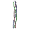
| ||||||||
|---|---|---|---|---|---|---|---|---|---|
| 1 |
| ||||||||
| Unit cell |
|
-Components
| #1: Protein | Mass: 12279.212 Da / Num. of mol.: 3 / Fragment: HR1 domain, UNP residues 45-155 Source method: isolated from a genetically manipulated source Source: (gene. exp.) Mus musculus (house mouse) / Gene: Ptrf / Production host:  Escherichia coli (E. coli) / Strain (production host): BL21 Rosetta / References: UniProt: O54724 Escherichia coli (E. coli) / Strain (production host): BL21 Rosetta / References: UniProt: O54724#2: Water | ChemComp-HOH / | Water Mass: 18.015 Da / Num. of mol.: 13 / Source method: isolated from a natural source / Formula: H2O Mass: 18.015 Da / Num. of mol.: 13 / Source method: isolated from a natural source / Formula: H2O |
|---|
-Experimental details
-Experiment
| Experiment | Method: X-RAY DIFFRACTION / Number of used crystals: 1 |
|---|
- Sample preparation
| Crystal | Density Matthews: 2.88 Å3/Da / Density % sol: 57.31 % |
|---|---|
| Crystal grow | Temperature: 298 K / Method: vapor diffusion, sitting drop / pH: 7.6 Details: 0.1M HEPES (pH 7.6), 30% MPD, VAPOR DIFFUSION, SITTING DROP, temperature 298K |
-Data collection
| Diffraction | Mean temperature: 100 K |
|---|---|
| Diffraction source | Source: SYNCHROTRON / Site: Australian Synchrotron  / Beamline: MX2 / Wavelength: 0.979 Å / Beamline: MX2 / Wavelength: 0.979 Å |
| Detector | Type: ADSC QUANTUM 315r / Detector: CCD |
| Radiation | Monochromator: Undulator / Protocol: MAD / Monochromatic (M) / Laue (L): M / Scattering type: x-ray |
| Radiation wavelength | Wavelength: 0.979 Å / Relative weight: 1 |
| Reflection | Resolution: 3→52.4 Å / Num. obs: 9020 / % possible obs: 99.7 % / Observed criterion σ(I): 5.4 / Redundancy: 8.3 % / Rmerge(I) obs: 0.071 / Net I/σ(I): 14.9 |
| Reflection shell | Resolution: 3→3 Å / Redundancy: 8.6 % / Rmerge(I) obs: 0.295 / Mean I/σ(I) obs: 5.4 / % possible all: 99.9 |
- Processing
| Software |
| ||||||||||||||||||||||||||||||||||||||||||||||||||||||||||||||||||||||||||||||||||||||||||||||||||||
|---|---|---|---|---|---|---|---|---|---|---|---|---|---|---|---|---|---|---|---|---|---|---|---|---|---|---|---|---|---|---|---|---|---|---|---|---|---|---|---|---|---|---|---|---|---|---|---|---|---|---|---|---|---|---|---|---|---|---|---|---|---|---|---|---|---|---|---|---|---|---|---|---|---|---|---|---|---|---|---|---|---|---|---|---|---|---|---|---|---|---|---|---|---|---|---|---|---|---|---|---|---|
| Refinement | Method to determine structure: molecular replacement/SAD / Resolution: 3→37.886 Å / SU ML: 0.39 / σ(F): 1.34 / Phase error: 32.51 / Stereochemistry target values: ML
| ||||||||||||||||||||||||||||||||||||||||||||||||||||||||||||||||||||||||||||||||||||||||||||||||||||
| Solvent computation | Shrinkage radii: 0.9 Å / VDW probe radii: 1.11 Å / Solvent model: FLAT BULK SOLVENT MODEL | ||||||||||||||||||||||||||||||||||||||||||||||||||||||||||||||||||||||||||||||||||||||||||||||||||||
| Refinement step | Cycle: LAST / Resolution: 3→37.886 Å
| ||||||||||||||||||||||||||||||||||||||||||||||||||||||||||||||||||||||||||||||||||||||||||||||||||||
| Refine LS restraints |
| ||||||||||||||||||||||||||||||||||||||||||||||||||||||||||||||||||||||||||||||||||||||||||||||||||||
| LS refinement shell |
| ||||||||||||||||||||||||||||||||||||||||||||||||||||||||||||||||||||||||||||||||||||||||||||||||||||
| Refinement TLS params. | Method: refined / Refine-ID: X-RAY DIFFRACTION
| ||||||||||||||||||||||||||||||||||||||||||||||||||||||||||||||||||||||||||||||||||||||||||||||||||||
| Refinement TLS group |
|
