 Movie
Movie Controller


+ Open data

- Basic information
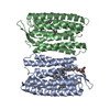
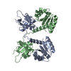
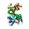
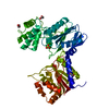
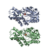
| Entry | Database: PDB / ID: 4qid | ||||||
|---|---|---|---|---|---|---|---|
| Title | Crystal structure of Haloquadratum walsbyi bacteriorhodopsin | ||||||
 Components Components | Bacteriorhodopsin-I | ||||||
 Keywords Keywords | MEMBRANE PROTEIN / Bacteriorhodopsin / proton pump / membrane | ||||||
| Function / homology |  Function and homology informationphotoreceptor activity / phototransduction / proton transmembrane transport / monoatomic ion channel activity / plasma membrane Function and homology informationphotoreceptor activity / phototransduction / proton transmembrane transport / monoatomic ion channel activity / plasma membraneSimilarity search - Function | ||||||
| Biological species |  Haloquadratum walsbyi (archaea) Haloquadratum walsbyi (archaea) | ||||||
| Method | X-RAY DIFFRACTION / SYNCHROTRON / MOLECULAR REPLACEMENT / Resolution: 2.57 Å | ||||||
 Authors Authors | Wang, A.H.J. / Hsu, M.F. / Yang, C.S. / Fu, H.Y. | ||||||
 Citation Citation | Journal: To be Published Title: An acid-tolerant light-driven proton pump Authors: Wang, A.H.J. / Hsu, M.F. / Yang, C.S. / Fu, H.Y. / Cai, C.J. / Yi, H.P. | ||||||
| History |
|
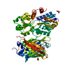
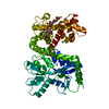
- Structure visualization
| Structure viewer | Molecule: MolmilJmol/JSmol |
|---|
- Downloads & links
-Download
| PDBx/mmCIF format | 4qid.cif.gz | 98 KB | Display | PDBx/mmCIF format |
|---|---|---|---|---|
| PDB format | pdb4qid.ent.gz | 81 KB | Display | PDB format |
| PDBx/mmJSON format | 4qid.json.gz | Tree view | PDBx/mmJSON format | |
| Others |  Other downloads Other downloads |
-Validation report
| Arichive directory | https://data.pdbj.org/pub/pdb/validation_reports/qi/4qidftp://data.pdbj.org/pub/pdb/validation_reports/qi/4qid | HTTPS FTP |
|---|
-Related structure data
| Related structure data | |
|---|---|
| Similar structure data |


-Links
 PDBj
PDBj


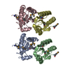
- Assembly
| Deposited unit | 
| ||||||||
|---|---|---|---|---|---|---|---|---|---|
| 1 |
| ||||||||
| Unit cell |
|
-Components
| #1: Protein | Mass: 28490.088 Da / Num. of mol.: 2 Source method: isolated from a genetically manipulated source Source: (gene. exp.) Haloquadratum walsbyi (archaea) / Strain: DSM 16790 / HBSQ001 / Gene: bop1, bopI, HQ_1014A / Plasmid: pET21d / Production host:  Escherichia coli (E. coli) / Strain (production host): C43 (DE3) / References: UniProt: Q18DH8 Escherichia coli (E. coli) / Strain (production host): C43 (DE3) / References: UniProt: Q18DH8#2: Chemical | Retinal  Mass: 284.436 Da / Num. of mol.: 2 / Source method: obtained synthetically / Formula: C20H28O Mass: 284.436 Da / Num. of mol.: 2 / Source method: obtained synthetically / Formula: C20H28O#3: Chemical | Acetate  Mass: 59.044 Da / Num. of mol.: 2 / Source method: obtained synthetically / Formula: C2H3O2 Mass: 59.044 Da / Num. of mol.: 2 / Source method: obtained synthetically / Formula: C2H3O2#4: Chemical | ChemComp-MPG / [( |   Mass: 356.540 Da / Num. of mol.: 1 / Source method: obtained synthetically / Formula: C21H40O4 Mass: 356.540 Da / Num. of mol.: 1 / Source method: obtained synthetically / Formula: C21H40O4#5: Water | ChemComp-HOH / | Water Mass: 18.015 Da / Num. of mol.: 102 / Source method: isolated from a natural source / Formula: H2O Mass: 18.015 Da / Num. of mol.: 102 / Source method: isolated from a natural source / Formula: H2O |
|---|
-Experimental details
-Experiment
| Experiment | Method: X-RAY DIFFRACTION / Number of used crystals: 1 |
|---|
- Sample preparation
| Crystal | Density Matthews: 1.89 Å3/Da / Density % sol: 34.92 % |
|---|---|
| Crystal grow | Temperature: 293 K / Method: lipidic cubic phase / pH: 4 Details: ammonium sulfate, NaCl, sodium acetate, PEG 200, pH 4.0, LIPID CUBIC PHASE, temperature 293.0K |
-Data collection
| Diffraction | Mean temperature: 110 K |
|---|---|
| Diffraction source | Source: SYNCHROTRON / Site: SPring-8  / Beamline: BL44XU / Wavelength: 0.9 Å / Beamline: BL44XU / Wavelength: 0.9 Å |
| Detector | Type: RAYONIX MX-225 / Detector: CCD / Date: Apr 20, 2013 |
| Radiation | Monochromator: double-crystal / Protocol: SINGLE WAVELENGTH / Monochromatic (M) / Laue (L): M / Scattering type: x-ray |
| Radiation wavelength | Wavelength: 0.9 Å / Relative weight: 1 |
| Reflection | Resolution: 2.57→30 Å / Num. obs: 48309 / % possible obs: 97.8 % / Observed criterion σ(F): 0 / Observed criterion σ(I): -3 / Redundancy: 3.5 % / Rmerge(I) obs: 0.168 / Rsym value: 0.168 / Net I/σ(I): 8.212 |
| Reflection shell | Resolution: 2.58→2.67 Å / Redundancy: 3.1 % / Rmerge(I) obs: 0.791 / Mean I/σ(I) obs: 2.016 / Rsym value: 0.791 / % possible all: 98 |
- Processing
| Software |
| ||||||||||||||||||||||||||||||||||||||||||||||||||||||||||||
|---|---|---|---|---|---|---|---|---|---|---|---|---|---|---|---|---|---|---|---|---|---|---|---|---|---|---|---|---|---|---|---|---|---|---|---|---|---|---|---|---|---|---|---|---|---|---|---|---|---|---|---|---|---|---|---|---|---|---|---|---|---|
| Refinement | Method to determine structure: MOLECULAR REPLACEMENT / Resolution: 2.57→28.87 Å / Cor.coef. Fo:Fc: 0.94 / Cor.coef. Fo:Fc free: 0.928 / SU B: 8.435 / SU ML: 0.19 / Cross valid method: THROUGHOUT / ESU R: 0.717 / ESU R Free: 0.32 / Stereochemistry target values: MAXIMUM LIKELIHOOD / Details: HYDROGENS HAVE BEEN ADDED IN THE RIDING POSITIONS
| ||||||||||||||||||||||||||||||||||||||||||||||||||||||||||||
| Solvent computation | Ion probe radii: 0.8 Å / Shrinkage radii: 0.8 Å / VDW probe radii: 1.2 Å / Solvent model: MASK | ||||||||||||||||||||||||||||||||||||||||||||||||||||||||||||
| Displacement parameters | Biso mean: 48.988 Å2
| ||||||||||||||||||||||||||||||||||||||||||||||||||||||||||||
| Refinement step | Cycle: LAST / Resolution: 2.57→28.87 Å
| ||||||||||||||||||||||||||||||||||||||||||||||||||||||||||||
| Refine LS restraints |
| ||||||||||||||||||||||||||||||||||||||||||||||||||||||||||||
| LS refinement shell | Resolution: 2.568→2.635 Å / Total num. of bins used: 20
|
