- PDB-4q4l: Crystal structure of an ATP synthase subunit beta 1 (F1-B1) from ... -
+
Open data
ID or keywords:
Loading...
-
Basic information
Entry
Database: PDB / ID: 4q4l
Title
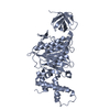
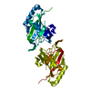
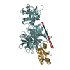
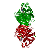
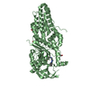
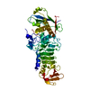
Crystal structure of an ATP synthase subunit beta 1 (F1-B1) from Burkholderia thailandensis
Components
ATP synthase subunit beta 1
Keywords
HYDROLASE / Structural Genomics / NIAID / National Institute of Allergy and Infectious Diseases / Seattle Structural Genomics Center for Infectious Disease / SSGCID / ATP-binding / metal ion binding / heterooligomeric protein complex / multidomain protein
Function / homology
Function and homology information
proton-transporting ATP synthase complex, catalytic core F(1) / H+-transporting two-sector ATPase / proton-transporting ATPase activity, rotational mechanism / proton-transporting ATP synthase activity, rotational mechanism / ATP binding / plasma membrane Similarity search - Function
Thrombin, subunit H - #170 / Bovine Mitochondrial F1-ATPase, ATP Synthase Beta Chain; Chain D, domain3 / Bovine Mitochondrial F1-atpase; Atp Synthase Beta Chain; Chain D, domain 3 / ATP synthase, F1 complex, beta subunit / ATPase, F1/V1 complex, beta/alpha subunit, C-terminal / ATPase, F1/V1/A1 complex, alpha/beta subunit, N-terminal domain superfamily / ATPase, F1/V1/A1 complex, alpha/beta subunit, N-terminal domain / ATP synthase alpha/beta family, beta-barrel domain / ATPase, alpha/beta subunit, nucleotide-binding domain, active site / ATP synthase alpha and beta subunits signature. ...Thrombin, subunit H - #170 / Bovine Mitochondrial F1-ATPase, ATP Synthase Beta Chain; Chain D, domain3 / Bovine Mitochondrial F1-atpase; Atp Synthase Beta Chain; Chain D, domain 3 / ATP synthase, F1 complex, beta subunit / ATPase, F1/V1 complex, beta/alpha subunit, C-terminal / ATPase, F1/V1/A1 complex, alpha/beta subunit, N-terminal domain superfamily / ATPase, F1/V1/A1 complex, alpha/beta subunit, N-terminal domain / ATP synthase alpha/beta family, beta-barrel domain / ATPase, alpha/beta subunit, nucleotide-binding domain, active site / ATP synthase alpha and beta subunits signature. / ATPase, F1/V1/A1 complex, alpha/beta subunit, nucleotide-binding domain / ATP synthase alpha/beta family, nucleotide-binding domain / Thrombin, subunit H / P-loop containing nucleotide triphosphate hydrolases / ATPases associated with a variety of cellular activities / AAA+ ATPase domain / Beta Barrel / P-loop containing nucleoside triphosphate hydrolase / Rossmann fold / Orthogonal Bundle / 3-Layer(aba) Sandwich / Mainly Beta / Mainly Alpha / Alpha Beta Similarity search - Domain/homology
Mass: 18.015 Da / Num. of mol.: 110 / Source method: isolated from a natural source / Formula: H2O
-
Experimental details
-
Experiment
Experiment
Method: X-RAY DIFFRACTION / Number of used crystals: 1
-
Sample preparation
Crystal
Density Matthews: 2.68 Å3/Da / Density % sol: 54.09 %
Crystal grow
Temperature: 289 K / Method: vapor diffusion, sitting drop / pH: 8 Details: ButhA.17369.c.A1.PW35952 at 19.9 mg/mL with 1 mM MnCl2 and 1 mM ATPgS against PACT screen condition B5, 0.1 M MIB buffer pH 8.0, 25% PEG 1500 with 20% ethylene glycol as cryo-protectant, ...Details: ButhA.17369.c.A1.PW35952 at 19.9 mg/mL with 1 mM MnCl2 and 1 mM ATPgS against PACT screen condition B5, 0.1 M MIB buffer pH 8.0, 25% PEG 1500 with 20% ethylene glycol as cryo-protectant, crystal tracking ID 242856b5, unique tracking ID ppm0-6, VAPOR DIFFUSION, SITTING DROP, temperature 289K
Resolution: 2.2→50 Å / Cor.coef. Fo:Fc: 0.957 / Cor.coef. Fo:Fc free: 0.943 / WRfactor Rfree: 0.2327 / WRfactor Rwork: 0.1901 / FOM work R set: 0.8186 / SU B: 11.525 / SU ML: 0.143 / SU R Cruickshank DPI: 0.2048 / SU Rfree: 0.1814 / Cross valid method: THROUGHOUT / σ(F): 0 / ESU R: 0.205 / ESU R Free: 0.181 / Stereochemistry target values: MAXIMUM LIKELIHOOD Details: U VALUES : WITH TLS ADDED HYDROGENS HAVE BEEN ADDED IN THE RIDING POSITIONS
Rfactor
Num. reflection
% reflection
Selection details
Rfree
0.2361
1462
4.9 %
RANDOM
Rwork
0.1939
-
-
-
obs
0.1959
29696
99.87 %
-
Solvent computation
Ion probe radii: 0.8 Å / Shrinkage radii: 0.8 Å / VDW probe radii: 1.2 Å / Solvent model: MASK
In the structure databanks used in Yorodumi, some data are registered as the other names, "COVID-19 virus" and "2019-nCoV". Here are the details of the virus and the list of structure data.
Jan 31, 2019. EMDB accession codes are about to change! (news from PDBe EMDB page)
EMDB accession codes are about to change! (news from PDBe EMDB page)
The allocation of 4 digits for EMDB accession codes will soon come to an end. Whilst these codes will remain in use, new EMDB accession codes will include an additional digit and will expand incrementally as the available range of codes is exhausted. The current 4-digit format prefixed with “EMD-” (i.e. EMD-XXXX) will advance to a 5-digit format (i.e. EMD-XXXXX), and so on. It is currently estimated that the 4-digit codes will be depleted around Spring 2019, at which point the 5-digit format will come into force.
The EM Navigator/Yorodumi systems omit the EMD- prefix.
Related info.:Q: What is EMD? / ID/Accession-code notation in Yorodumi/EM Navigator
Yorodumi is a browser for structure data from EMDB, PDB, SASBDB, etc.
This page is also the successor to EM Navigator detail page, and also detail information page/front-end page for Omokage search.
The word "yorodu" (or yorozu) is an old Japanese word meaning "ten thousand". "mi" (miru) is to see.
Related info.:EMDB / PDB / SASBDB / Comparison of 3 databanks / Yorodumi Search / Aug 31, 2016. New EM Navigator & Yorodumi / Yorodumi Papers / Jmol/JSmol / Function and homology information / Changes in new EM Navigator and Yorodumi
 Movie
Movie Controller

 Yorodumi
 Open data
 Basic information
 Components
 Keywords
 Function and homology information
 Authors
 Citation
 Structure visualization
 Downloads & links
 Other downloads
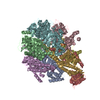


 PDBj
PDBj


 Assembly

 Mass: 22.990 Da / Num. of mol.: 1 / Source method: obtained synthetically / Formula: Na
 Mass: 18.015 Da / Num. of mol.: 110 / Source method: isolated from a natural source / Formula: H2O
 Sample preparation
 / Beamline: 21-ID-G / Wavelength: 0.97856 Å
 Processing