 Movie
Movie Controller


+ Open data

- Basic information
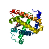
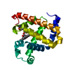
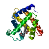
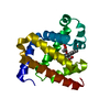
| Entry | Database: PDB / ID: 4q2s | ||||||
|---|---|---|---|---|---|---|---|
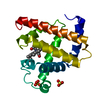
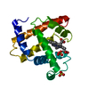
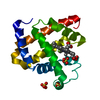
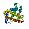
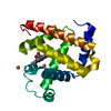
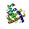
| Title | Crystal Structure of S. pombe Pdc1 Ge1 Domain | ||||||
 Components Components | PDC1 GE1 DOMAIN | ||||||
 Keywords Keywords |  RNA BINDING PROTEIN / Ge1 domain / P-body assembly RNA BINDING PROTEIN / Ge1 domain / P-body assembly | ||||||
| Function / homology | deadenylation-independent decapping of nuclear-transcribed mRNA / P-body assembly / P-body / cytoplasmic stress granule / molecular adaptor activity / WD40-repeat-containing domain superfamily / WD40/YVTN repeat-like-containing domain superfamily / cytoplasm / Uncharacterized protein C20G4.08 Function and homology information Function and homology information | ||||||
| Biological species |  Schizosaccharomyces pombe (fission yeast) Schizosaccharomyces pombe (fission yeast) | ||||||
| Method | X-RAY DIFFRACTION / SYNCHROTRON / after obtaining initial molecular replacement model by S-SAD / Resolution: 1.35 Å | ||||||
 Authors Authors | Noeldeke, E.R. / Neu, A. / Zocher, G. / Sprangers, R. | ||||||
 Citation Citation | Journal: Angew.Chem.Int.Ed.Engl. / Year: 2014 Title: In vitro reconstitution of a cellular phase-transition process that involves the mRNA decapping machinery. Authors: Fromm, S.A. / Kamenz, J. / Noldeke, E.R. / Neu, A. / Zocher, G. / Sprangers, R. | ||||||
| History |
|
- Structure visualization
| Structure viewer | Molecule: MolmilJmol/JSmol |
|---|
- Downloads & links
-Download
| PDBx/mmCIF format | 4q2s.cif.gz | 69.2 KB | Display | PDBx/mmCIF format |
|---|---|---|---|---|
| PDB format | pdb4q2s.ent.gz | 51.2 KB | Display | PDB format |
| PDBx/mmJSON format | 4q2s.json.gz | Tree view | PDBx/mmJSON format | |
| Others |  Other downloads Other downloads |
-Validation report
| Arichive directory | https://data.pdbj.org/pub/pdb/validation_reports/q2/4q2sftp://data.pdbj.org/pub/pdb/validation_reports/q2/4q2s | HTTPS FTP |
|---|
-Related structure data
| Similar structure data |
|---|
-Links
 PDBj
PDBj- Assembly
| Deposited unit | 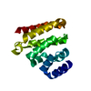
| ||||||||
|---|---|---|---|---|---|---|---|---|---|
| 1 |
| ||||||||
| Unit cell |
|
-Components
| #1: Protein | Mass: 16241.898 Da / Num. of mol.: 1 / Fragment: unp residues 932-1070 Source method: isolated from a genetically manipulated source Source: (gene. exp.) Schizosaccharomyces pombe (fission yeast)Strain: 972 / ATCC 24843 / Gene: SPAC20G4.08, SPAC4F10.01 / Production host:  Escherichia coli (E. coli) / References: UniProt: O13892 Escherichia coli (E. coli) / References: UniProt: O13892 |
|---|---|
| #2: Water | ChemComp-HOH / Water Mass: 18.015 Da / Num. of mol.: 70 / Source method: isolated from a natural source / Formula: H2O Mass: 18.015 Da / Num. of mol.: 70 / Source method: isolated from a natural source / Formula: H2O |
-Experimental details
-Experiment
| Experiment | Method: X-RAY DIFFRACTION / Number of used crystals: 1 |
|---|
- Sample preparation
| Crystal | Density Matthews: 2.07 Å3/Da / Density % sol: 40.59 % |
|---|---|
| Crystal grow | Temperature: 298 K / Method: vapor diffusion, sitting drop / pH: 7.5 Details: 0.2 M Ammonium Acetate, 0.1 M HEPES, 25% (w/v) PEG 3350, pH 7.5, VAPOR DIFFUSION, SITTING DROP, temperature 298K |
-Data collection
| Diffraction | Mean temperature: 100 K |
|---|---|
| Diffraction source | Source: SYNCHROTRON / Site: SLS  / Beamline: X10SA / Wavelength: 1 Å / Beamline: X10SA / Wavelength: 1 Å |
| Detector | Type: PSI PILATUS 6M / Detector: PIXEL / Date: Aug 19, 2013 |
| Radiation | Monochromator: Si(111) / Protocol: SINGLE WAVELENGTH / Monochromatic (M) / Laue (L): M / Scattering type: x-ray |
| Radiation wavelength | Wavelength: 1 Å / Relative weight: 1 |
| Reflection | Resolution: 1.35→40.4 Å / Num. all: 30393 / Num. obs: 30287 / % possible obs: 99.7 % / Observed criterion σ(I): -2 |
| Reflection shell | Resolution: 1.35→1.39 Å / % possible all: 100 |
- Processing
| Software |
| ||||||||||||||||||||||||||||||||||||||||||||||||||||||||||||||||||||||||||||||||||||||||||||||||||||||||||||||||||||||||
|---|---|---|---|---|---|---|---|---|---|---|---|---|---|---|---|---|---|---|---|---|---|---|---|---|---|---|---|---|---|---|---|---|---|---|---|---|---|---|---|---|---|---|---|---|---|---|---|---|---|---|---|---|---|---|---|---|---|---|---|---|---|---|---|---|---|---|---|---|---|---|---|---|---|---|---|---|---|---|---|---|---|---|---|---|---|---|---|---|---|---|---|---|---|---|---|---|---|---|---|---|---|---|---|---|---|---|---|---|---|---|---|---|---|---|---|---|---|---|---|---|---|
| Refinement | Method to determine structure: after obtaining initial molecular replacement model by S-SAD Resolution: 1.35→40.44 Å / Cor.coef. Fo:Fc: 0.961 / Cor.coef. Fo:Fc free: 0.963 / SU B: 2.516 / SU ML: 0.046 / Cross valid method: THROUGHOUT / ESU R: 0.066 / ESU R Free: 0.06 / Stereochemistry target values: MAXIMUM LIKELIHOOD / Details: HYDROGENS HAVE BEEN ADDED IN THE RIDING POSITIONS
| ||||||||||||||||||||||||||||||||||||||||||||||||||||||||||||||||||||||||||||||||||||||||||||||||||||||||||||||||||||||||
| Solvent computation | Ion probe radii: 0.8 Å / Shrinkage radii: 0.8 Å / VDW probe radii: 1.2 Å / Solvent model: MASK | ||||||||||||||||||||||||||||||||||||||||||||||||||||||||||||||||||||||||||||||||||||||||||||||||||||||||||||||||||||||||
| Displacement parameters | Biso mean: 19.888 Å2
| ||||||||||||||||||||||||||||||||||||||||||||||||||||||||||||||||||||||||||||||||||||||||||||||||||||||||||||||||||||||||
| Refinement step | Cycle: LAST / Resolution: 1.35→40.44 Å
| ||||||||||||||||||||||||||||||||||||||||||||||||||||||||||||||||||||||||||||||||||||||||||||||||||||||||||||||||||||||||
| Refine LS restraints |
| ||||||||||||||||||||||||||||||||||||||||||||||||||||||||||||||||||||||||||||||||||||||||||||||||||||||||||||||||||||||||
| LS refinement shell | Resolution: 1.35→1.385 Å / Total num. of bins used: 20
|
