 Movie
Movie Controller

[English] 日本語
 Yorodumi
- PDB-4pu3: Shewanella oneidensis MR-1 Toxin Antitoxin System HipA, HipB and ... -
+ Open data

- Basic information
| Entry | Database: PDB / ID: 4pu3 | ||||||
|---|---|---|---|---|---|---|---|
| Title | Shewanella oneidensis MR-1 Toxin Antitoxin System HipA, HipB and its operator DNA complex (space group P212121) | ||||||
 Components Components |
| ||||||
 Keywords Keywords | TOXIN/ANTITOXIN/DNA / Toxin Antitoxin System / TOXIN-ANTITOXIN-DNA complex | ||||||
| Function / homology |  Function and homology information Function and homology information non-specific serine/threonine protein kinase / phosphorylation / protein serine kinase activity / protein serine/threonine kinase activity / DNA binding / ATP binding / cytosol non-specific serine/threonine protein kinase / phosphorylation / protein serine kinase activity / protein serine/threonine kinase activity / DNA binding / ATP binding / cytosolSimilarity search - Function | ||||||
| Biological species |  Shewanella oneidensis (bacteria) Shewanella oneidensis (bacteria) | ||||||
| Method | X-RAY DIFFRACTION / SYNCHROTRON / MOLECULAR REPLACEMENT / Resolution: 3.39 Å | ||||||
 Authors Authors | Wen, Y. / Behiels, E. / Felix, J. / Elegheert, J. / Vergauwen, B. / Devreese, B. / Savvides, S. | ||||||
 Citation Citation | Journal: Nucleic Acids Res. / Year: 2014 Title: The bacterial antitoxin HipB establishes a ternary complex with operator DNA and phosphorylated toxin HipA to regulate bacterial persistence. Authors: Wen, Y. / Behiels, E. / Felix, J. / Elegheert, J. / Vergauwen, B. / Devreese, B. / Savvides, S.N. | ||||||
| History |
|
- Structure visualization
| Structure viewer | Molecule: MolmilJmol/JSmol |
|---|
- Downloads & links
-Download
| PDBx/mmCIF format | 4pu3.cif.gz | 413.9 KB | Display | PDBx/mmCIF format |
|---|---|---|---|---|
| PDB format | pdb4pu3.ent.gz | 337.2 KB | Display | PDB format |
| PDBx/mmJSON format | 4pu3.json.gz | Tree view | PDBx/mmJSON format | |
| Others |  Other downloads Other downloads |
-Validation report
| Arichive directory | https://data.pdbj.org/pub/pdb/validation_reports/pu/4pu3ftp://data.pdbj.org/pub/pdb/validation_reports/pu/4pu3 | HTTPS FTP |
|---|
-Related structure data
| Related structure data | 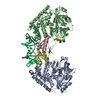 4pu4C  4pu5C  4pu7C  4pu8C  3dnvS C: citing same article ( S: Starting model for refinement |
|---|---|
| Similar structure data |


-Links
 PDBj
PDBj


- Assembly
| Deposited unit | 
| |||||||||||||||||||||||||||||||||||||||||||||||||||||||||||||||||||
|---|---|---|---|---|---|---|---|---|---|---|---|---|---|---|---|---|---|---|---|---|---|---|---|---|---|---|---|---|---|---|---|---|---|---|---|---|---|---|---|---|---|---|---|---|---|---|---|---|---|---|---|---|---|---|---|---|---|---|---|---|---|---|---|---|---|---|---|---|
| 1 |
| |||||||||||||||||||||||||||||||||||||||||||||||||||||||||||||||||||
| Unit cell |
| |||||||||||||||||||||||||||||||||||||||||||||||||||||||||||||||||||
| Noncrystallographic symmetry (NCS) | NCS domain:
NCS domain segments:
NCS ensembles :
|
-Components
| #1: Protein | Mass: 51167.391 Da / Num. of mol.: 2 Source method: isolated from a genetically manipulated source Source: (gene. exp.) Shewanella oneidensis (bacteria) / Strain: MR-1 / Gene: SO_0706 / Production host: Escherichia coli (E. coli) / Strain (production host): BL21(DE3) / References: UniProt: Q8EIX3#2: Protein | / HipBMass: 12951.791 Da / Num. of mol.: 2 Source method: isolated from a genetically manipulated source Source: (gene. exp.) Shewanella oneidensis (bacteria) / Strain: MR-1 / Gene: SO_0705 / Production host: Escherichia coli (E. coli) / References: UniProt: Q8EIX4#3: DNA chain | | Mass: 8036.256 Da / Num. of mol.: 1 / Source method: obtained synthetically / Details: strand 1 / Source: (synth.) Shewanella oneidensis (bacteria)#4: DNA chain | | Mass: 7933.134 Da / Num. of mol.: 1 / Source method: obtained synthetically / Details: strand 2 / Source: (synth.) Shewanella oneidensis (bacteria) |
|---|
-Experimental details
-Experiment
| Experiment | Method: X-RAY DIFFRACTION / Number of used crystals: 1 |
|---|
- Sample preparation
| Crystal | Density Matthews: 2.33 Å3/Da / Density % sol: 47.22 % |
|---|---|
| Crystal grow | Temperature: 293 K / Method: vapor diffusion / pH: 7.5 Details: 0.2 M calcium acetate hydrate, pH 7.5, 20% PEG3350, VAPOR DIFFUSION, temperature 293K |
-Data collection
| Diffraction |
| ||||||||||||||||||
|---|---|---|---|---|---|---|---|---|---|---|---|---|---|---|---|---|---|---|---|
| Diffraction source |
| ||||||||||||||||||
| Detector |
| ||||||||||||||||||
| Radiation |
| ||||||||||||||||||
| Radiation wavelength | Wavelength: 1 Å / Relative weight: 1 | ||||||||||||||||||
| Reflection | Resolution: 3.39→49.39 Å / Num. all: 19211 / Num. obs: 19211 / % possible obs: 98.93 % / Observed criterion σ(I): 1.81 | ||||||||||||||||||
| Reflection shell | Highest resolution: 3.39 Å |


- Processing
| Software |
| ||||||||||||||||||||||||||||||||||||||||||||||||||||||||
|---|---|---|---|---|---|---|---|---|---|---|---|---|---|---|---|---|---|---|---|---|---|---|---|---|---|---|---|---|---|---|---|---|---|---|---|---|---|---|---|---|---|---|---|---|---|---|---|---|---|---|---|---|---|---|---|---|---|
| Refinement | Method to determine structure: MOLECULAR REPLACEMENT Starting model: PDB ENTRY 3DNV Resolution: 3.39→49.381 Å / SU ML: 0.57 / σ(F): 1.99 / Phase error: 35.22 / Stereochemistry target values: ML
| ||||||||||||||||||||||||||||||||||||||||||||||||||||||||
| Solvent computation | Shrinkage radii: 0.9 Å / VDW probe radii: 1.11 Å / Solvent model: FLAT BULK SOLVENT MODEL | ||||||||||||||||||||||||||||||||||||||||||||||||||||||||
| Refinement step | Cycle: LAST / Resolution: 3.39→49.381 Å
| ||||||||||||||||||||||||||||||||||||||||||||||||||||||||
| Refine LS restraints |
| ||||||||||||||||||||||||||||||||||||||||||||||||||||||||
| Refine LS restraints NCS |
| ||||||||||||||||||||||||||||||||||||||||||||||||||||||||
| LS refinement shell |
|
