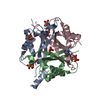
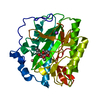
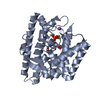
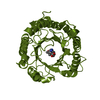
Entry Database : PDB / ID : 4ozjTitle GlnK2 from Haloferax mediterranei complexed with ADP Nitrogen regulatory protein P-II Keywords / / / / / / / Function / homology Function Domain/homology Component
/ / / / / / / / / / / / / / / / / / / / Biological species Haloferax mediterranei (archaea)Method / / Resolution : 1.45 Å Authors Palanca, C. / Pedro-Roig, L. / Llacer, J.L. / Camacho, M. / Bonete, M.J. / Rubio, V. Funding support Organization Grant number Country Spanish government BFU2011-30407 Spanish government BIO2008_00082 Valencian government Prometeo 2009/51
Journal : Febs J. / Year : 2014Title : The structure of a PII signaling protein from a halophilic archaeon reveals novel traits and high-salt adaptations.Authors : Palanca, C. / Pedro-Roig, L. / Llacer, J.L. / Camacho, M. / Bonete, M.J. / Rubio, V. History Deposition Feb 17, 2014 Deposition site / Processing site Revision 1.0 Jul 2, 2014 Provider / Type Revision 1.1 Oct 1, 2014 Group Revision 1.2 Dec 27, 2023 Group Data collection / Database references ... Data collection / Database references / Derived calculations / Other / Refinement description / Source and taxonomy Category chem_comp_atom / chem_comp_bond ... chem_comp_atom / chem_comp_bond / database_2 / diffrn_source / entity_src_gen / pdbx_database_status / pdbx_struct_assembly / pdbx_struct_assembly_prop / pdbx_struct_oper_list / refine_hist Item _database_2.pdbx_DOI / _database_2.pdbx_database_accession ... _database_2.pdbx_DOI / _database_2.pdbx_database_accession / _diffrn_source.pdbx_synchrotron_site / _entity_src_gen.pdbx_alt_source_flag / _pdbx_database_status.pdb_format_compatible / _pdbx_struct_assembly.oligomeric_details / _pdbx_struct_assembly_prop.type / _pdbx_struct_assembly_prop.value / _pdbx_struct_oper_list.symmetry_operation / _refine_hist.number_atoms_solvent / _refine_hist.number_atoms_total / _refine_hist.pdbx_number_atoms_ligand / _refine_hist.pdbx_number_atoms_nucleic_acid / _refine_hist.pdbx_number_atoms_protein
Show all Show less
 Movie
Movie Controller


 Open data
 Basic information
 Components
 Keywords
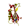
 SIGNALING PROTEIN / GlnK / PII / GlnB /
  Function and homology information
 Authors
 Spain, 3items
  Citation
 Structure visualization
 Downloads & links
 Other downloads


 PDBj
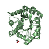
PDBj Assembly


 Mass: 427.201 Da / Num. of mol.: 1 / Source method: obtained synthetically / Formula: C10H15N5O10P2 / Comment: ADP, energy-carrying molecule*YM
 Mass: 18.015 Da / Num. of mol.: 72 / Source method: isolated from a natural source / Formula: H2O
 Sample preparation
 / Beamline: I04 / Wavelength: 0.9795 Å
 Processing